
INTRODUCTION
When restoring human matter with numerical concentrations, the following methods can be used:
1) Read the numbers of the series corresponding to the restored matter, written after the name of the matter;
2) Mentally pronounce the numbers of the series corresponding to the restored matter;
3) Look at the image or name of the matter and mentally pronounce the numbers of the corresponding row;
4) Imagine that you are between the numbers of the series corresponding to the matter being recreated, having a large size. The numbers between which you imagine yourself, you need to strive to perceive distinctly. The light from these numbers can reach you. The above actions can be performed with any numbers in the series.
5) Imagine that you are looking at a number series from above.
6) Represent a numerical series in the area that you are restoring. To do this, you need to use the image of matter given in this book, relating to the number series you are using.
7) Represent the numerical series between the image of matter and part of the mirror image given in this book that relate to the number series used.
8) By comparing the numbers of the series, you can determine the relationship between the various human matter that controls the norm. It is possible to restore matter using a series of other matter. In this case, when using the numerical series of the matter that is being restored, you can simultaneously or sequentially use the numerical series of other matter. From the numerical series of other matter, you can first concentrate on the numbers that coincide with the numbers of the series of the matter being restored. Then you can use the entire numerical series of other matter, mentally drawing a light beam from it that intersects the numerical series of the matter or the matter itself that you are restoring.
When perceiving a rapid restorative effect, you can determine the next point or area in the organism after the matter being restored itself, through which the restored matter is created. This next point or area will then be in other matter, through whose numerical series the restoration of the matter you have chosen is carried out. There may be many subsequent points or areas through which the creation of matter is carried out. The first point or area of creation of the selected matter is in the matter itself.
Having established the points or areas of creation of the restored matter by using numerical series, it is possible to restore the matter by concentrating on these points or areas. In this case, a spiritual state is established that corresponds to the restoration and norm of the selected matter.
Remembering and establishing the indicated state of the spirit, it is possible to restore the matter with the spirit, which is thus life-giving. Then it is possible to extend such spiritual action to all the matter of the organism, taking into account external events, and thereby achieve a spiritual state that corresponds to eternal development. In certain cases, depending on the angle of perception, different numerical series may correspond to the restored matter.
9) To speed up the restoration of human matter, gaps in numerical series can be perceived as gaps between words in a sentence. Then, for each numerical component of the entire series, separated by a space, you can consider the word that means the normal functioning of matter, to which the series corresponds. Creator the level that creates matter corresponding to the numerical series, and the matter of the whole organism can be perceived, trying to perceive such a word. Light, creating matter corresponding to the numerical series, spreads according to the laws of optics on all other matter of the human body and on the environment. Here you can understand why Some sensations and emotions are perceived as external.
This allows for more accurate recognition of where on the level
event management must act on the basis of the interaction of body tissues and where on the basis of interactions of the body's matter and the environment. This method of precise recognition allows more effectively manage events to the level of the normal state of the body's matter, regardless of any circumstances.
With this method, you simultaneously perceive the tissue of the body and the events surrounding the person. as if you were looking at what was described with your physical sight. And depending on the situation, you can take solution – how to act in the direction of eternal development. In some cases, you can take physical actions and in some periods to produce spiritual action to normalize events in the direction of eternal life.
This perception of yours develops your spirit, soul and physical body to the level at which creation of human matter occurs on a spiritual basis. Numbers allow us to obtain an accurate spiritual state, corresponding to the norm of human matter. To strengthen control, you can use the well-known, that is, knowledge from physics about the corpuscular-wave duality of matter, widely entrenched in the collective consciousness, according to which any object can exhibit both wave properties and the properties of a particle of matter.
Creating by concentrating on the number series of light waves corresponding to the norm of human matter, you create
normally functioning human matter.
All data in this book on the methods of restoring human matter using numerical concentrations can be use for preventive health purposes, for rejuvenation, if necessary to restore matter, and regardless of the initial data from which the matter is restored.
When using the specified In points 1 through 9 of the introduction of methods, the following can be taken into account:
1. For preventive purposes, it is advisable to carry out health improvement with the simultaneous distribution the effects of numerical concentrations on the future.
2. When rejuvenating, it is advisable to first consistently concentrate on the number series, located in the content, taking into account the task of eternal development, then concentrate on the matter that you
locally rejuvenate.
3. By restoring the body's matter, it is possible to produce numerical concentrations of successively different given in the book in the ways. In this case, it is possible to use both the numerical series corresponding to the reconstructed matter, as well as the numerical series of the area that includes the matter that you are restoring.
4. If it is necessary to restore matter after biological death, then one should concentrate on numbers first sequentially from left to right, then in reverse order from right to left.
The spiritual impulse that creates human matter allows for the expansion of recovery methods.
In restoring the matter of man, one must strive to develop the spiritual level to a state in which human matter is created and functions by spiritual action along with biological and event-based
principles.
Such a spiritual state, when implementing the methods of eternal development, should ensure complete restoration of human matter regardless of initial data and any circumstances.
HEMOPOIETIC SYSTEM AND
IMMUNE PROTECTION 219 648 317 918
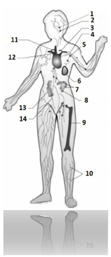
Fig. 1 Organs of the immune system: 214 317 498 817:
1 – adenoids 471 219 319 819
2 – palatine tonsils 428 641 478 591
3 – thoracic lymphatic duct 514 715 914 815
4 – subclavian vein 598 317 898 214
5 – lymph nodes 514 317 219 419
6 – spleen 548 711 918 321
7 – Peyer's patches 598 721 398 641
8 – small intestine 528 317 428 717
9 – red bone marrow 598 492 319 016
10 – lymphatic vessels 598 064 571 389
11 – right lymphatic duct 418 481 499 164
12 – thymus (thymus gland) 481 914 319 814
13 – Large intestine 591 488 898 217
14 – appendix 529 317 899 228
Central organs of hematopoiesis and
immune defense 416 489 319 641
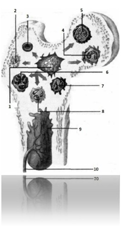
Fig. 2 Red bone marrow: 497 214 218 641:
1 – stem cell 451 618 719 481
2 – platelets 649 317 498 714
3 – erythrocyte 214 719 319 818
4 – monocyte 519 671 319 648
5 – lymphocyte 516 318 948 714
6 – basophil 319 648 719 814
7 – eosinophil 549 316 718 491
8 – neutrophil 467 589 891 648
9 – red (hematopoietic) bone marrow 497 214 218 641
10 – feeding artery 641 849 317 914
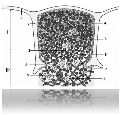
Fig. 3 Lobe of the thymus gland
(building) 519 713 219 498:
I – Cortex 519 648 219714
II – Medulla 319 498 516 814
1 – capsule 598 317 948 567
2 – partition 968 319 594 217
3 – macrophage 947 368 249 714
4 – epithelial cell of the cortex (cell –
nanny) 549 361 897 217
5 – thymocyte of the cortical layer 564 891 218 647
6 – epithelial cell of the medulla
574 016 217 498
7 – dendritic epithelial cell 598 671 390 149
8 – thymocyte of the medulla 531 649 174 061
9 – Hassal’s corpuscle 914 864 719 472
Peripheral organs of hematopoiesis and
immune defense 794 916 219 481
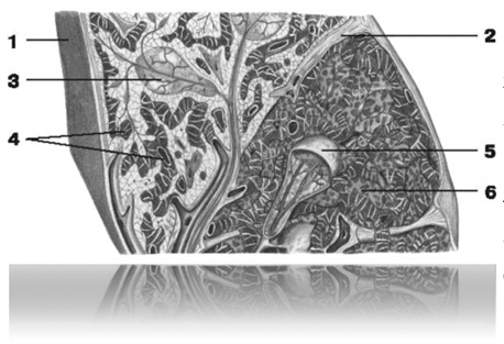
Fig. 4 Spleen (structure) 548 711 918 321:
1 – fibrous membrane 589 491 317 548
2 – trabecula of the spleen 317 489 896 104
3 – lymphoid follicles of the spleen
517 218 496 471
4 – venous sinuses 594 328 697 541
5 – white pulp 589 674 198 491
6 – red pulp 589 671 318 494
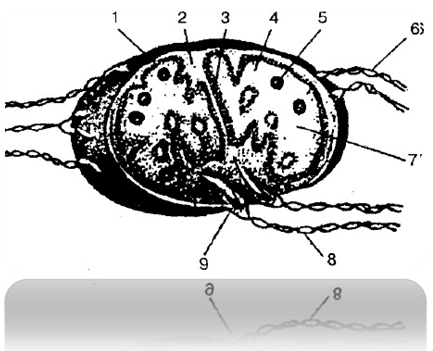
Fig. 5 Structure of the lymph node
591 148 319 888:
1 – capsule 519 848 718 949
2 – trabecula 518 716 918 317
3 – crossbar 898 749 219 317
4 – cortex 519 421 319 281
5 – follicles 898 715 984 355
6 – afferent lymphatic vessels
598 741 288 511
7 – brain substance 498 641 319 817
8 – efferent lymphatic vessels
512 789 319 489
9 – gate of the lymph node 598 681 724 918
Unified immune system of the mucous membranes
674 981 219 496
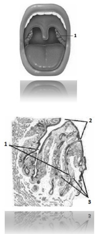
Fig. 6 Palatine tonsil
(location in the oral cavity)
514 218 319 671:
1 – palatine tonsil 514 218 319 671
Fig. 7 Palatine tonsil (structure)
514 218 319 671:
1 – gaps 894 316 548 917
2 – lymphatic follicles 598 641 317 214
3 – lacunae openings 349 548 671 214
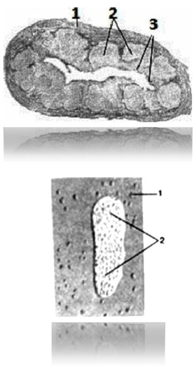
Fig. 8 Lymphoid nodules (in the wall of the appendix)
319 648 317 498:
1 – wall of appendix 217 214 218 641
2 – lymphoid nodules 319 648 317 498
3 – epithelial cover 218 491 016 648
Fig. 9 Lymphoid nodules and lymphoid plaque
in the wall of the small intestine 249 317 498 641:
1 – lymphoid nodules 548 547 198 678
2 – lymphoid plaque 589 641 948 581
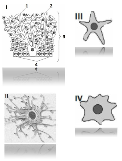
Fig. 10 Antigen presenting cells (APC)
598 647 319 591:
I – Langerhans cell 548 491 619 891
1 – Langerhans cell 548 491 619 891
2 – keratinocytes 516 891 719 478
3 – epidermis 598 718 889 888
4 – basement membrane 689 497 597 814
II – follicular dendritic cell
594 716 219 819
III – interdigital cell 548 217 319 471
IV – dendritic cell of the germinal centers
549 621 891 719
BLOOD CELLS 549681219717
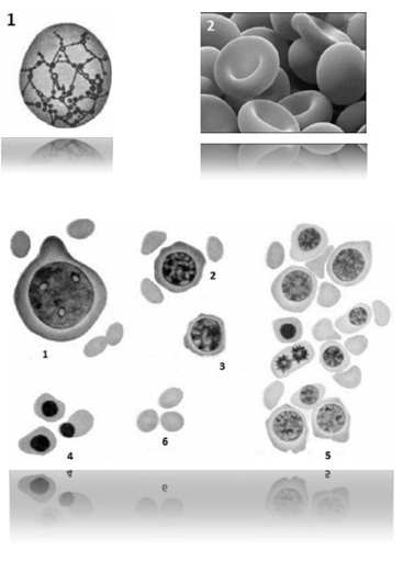
Fig. 11 Red blood cells: 467 198 219 814:
1 – blood reticulocyte (immature erythrocyte)
691 218 498 514
2 – red blood cells 467 198 219 814
Rice. 12 Erythrocytopoiesis: 489 617 519 318:
1 – erythroblast 589 649 218 717
2 – pronormocyte 218 719 814 798
3 – basophilic normocyte 496 198 217 248
4 – orthochromic normocyte 496 814 219 817
5 – polychromatophilic normocyte 598 694 197 281
6 – erythrocyte 214 719 319 818
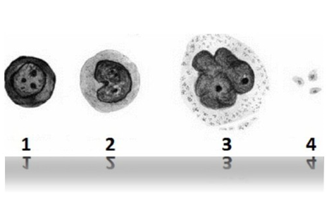
Fig. 13 Thrombocytopoiesis: 481 249 016 914:
1 – megakaryoblast 894 316 218 516
2 – promegakaryocyte 481 216 318 491
3 – megakaryocyte 471 218 694 271
4 – platelets 649 317 498 714
Thrombopoietin 890 648 019 312
Leukocytes 694 218 574 271
Agranulocytes 548 274 298 641
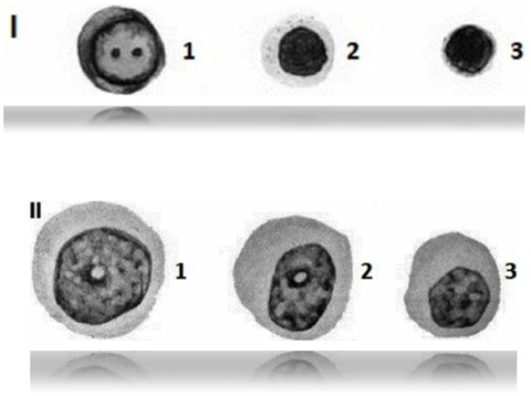
Fig. 14 Lymphocytopoiesis: 648 041 298 471:
I – B – lymphocytes 518 541 316 218
1 – B – lymphoblast 316 491 519 618
2 – immature B – lymphocyte 518 491 217 496
3 – mature B – lymphocyte 498 164 019 981
II – T – lymphocytes 467 198 964 217
1 – T – lymphoblast 316 514 816 274
2 – immature T – lymphocyte 619 754 218 316
3 – mature T – lymphocyte 689 148 686 217
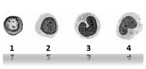
Fig. 15. Monocytopoiesis. Formation
monocytes and macrophages
496 514 218 471:
1 – monocytoblast 319 471 819 498
2 – promonocyte 619 814 516 714
3 – monocyte 519 671 319 648
4 – macrophage 947 368 249 714
Granulocytes 918 547 219 714
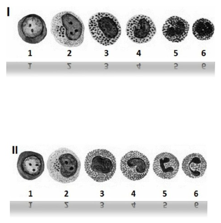
Fig. 16 Granulocytopoiesis: 214 617 218 549:
I – formation of basophilic granulocytes
584 316 318 491
1 – myeloblast 549 641 894 317
2 – promyelocyte 496 548 219 641
3 – myelocyte 517 219 498 641
4 – metamyelocyte 894 216 219 891
5 – band basophil 217 214 619 061
6 – segmented basophil 648 918 818 491
II – formation of eosinophilic granulocytes
496 549 718 546
1 – myeloblast 549 641 894 317
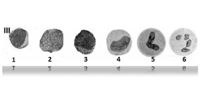
2 – promyelocyte 496 548 219 641
3 – myelocyte 517 219 498 641
4 – metamyelocyte 894 216 219 891
5 – band eosinophil 549 648 598 748
6 – segmented eosinophil 548 461 719 496
III – formation of neutrophilic granulocytes
564 581 498 641
1 – myeloblast 549 641 894 317
2 – promyelocyte 496 548 219 641
3 – myelocyte 517 219 498 641
4 – metamyelocyte 894 216 219 891
5 – band neutrophil 568 191 219 714
6 – segmented neutrophil 894 961 068 971
DENTOALVEOLAR SYSTEM 216 548 219 716
Bones of the facial part of the skull 219 715 819 815
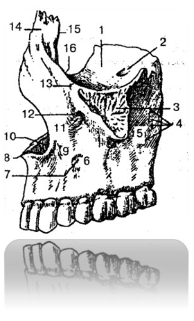
Fig. 17 Upper jaw (lateral view)
519 371 919 811
1 – orbital surface 398 216 718 226
2 – infraorbital groove 319 717 819 227
3 – zygomatic process 419 312 214 222
4 – alveolar openings 214 712 814 229
5 – temporal surface 538 722 918 222
6 – front surface 548 888 019 648
7 – canine fossa 539 717 819 317
8 – anterior nasal spine 529 513 919 813
9 – body of the upper jaw 548712 818 212
10 – nasal notch 548 716 298 444
11 – infraorbital canal 319 717 819 217
12 – infraorbital foramen 489 061 298 541
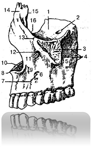
13 – zygomaticomaxillary suture 214 711 898 211
14 – frontal process 590 421 019 481
15 – lacrimal edge 548 884 918 888
16 – infraorbital margin 512 219 312 919
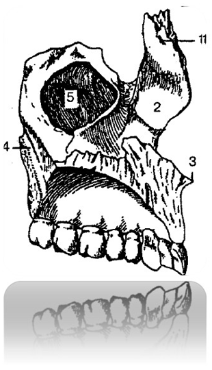
Fig. 18 Upper jaw (left) (view from
medial side) 421 718 911 328:
1 – frontal process 590 421 019 481
2 – nasal surface 598 648 319 711
3 – anterior nasal spine 529 513 919 813
4 – pterygopalatine groove 428 321 814 221
5 – maxillary sinus
519 321 814 471
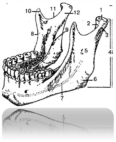
Fig. 19 Lower jaw 514 712 814 312:
1 – head of the lower jaw 548 321 848 721
2 – pterygoid fossa 519 317 919 007
3 – neck of the lower jaw 319 814 919 714
4, 5 – branches of the lower jaw 518 317 918 001
6 – angle of the lower jaw 548 219 289 008
7 – lower jaw canal 009 217 319 227
8 – temporal crest 418 317 228 227
9 – opening of the lower jaw 489 201 319 871
10 – coronoid process 528 317 918 228
11 – lower jaw notch 419 317 819 828
12 – condylar process 891 319 898 789
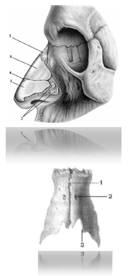
Fig. 20. Osteochondral skeleton of the external nose
948 547 219 641:
1 – nasal bone 518 314 818 214
2 – minor cartilages of the wings of the nose 916 814 219 618
3 – greater cartilage of the ala of the nose 719 316 219 494
4 – accessory nasal cartilage 989 617 318 641
5 – lateral cartilage 289 671 318 491
Fig. 21 Nasal bone 518 314 818 214:
1 – internasal suture 514 314 218 578
2 – opening of the nasal bone 316 581 314 891
3 – free edge 598 641 719 471
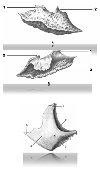
Fig. 22 Inferior nasal concha 478 218 918 217:
A - view from the outside
B - view from inside
1 – lacrimal process 548 671 219 491
2 – ethmoid process 491 897 319 648
3 – maxillary process 589 671 918 491
Fig. 23 Zygomatic bone 899 817 818 317:
1 – frontal process 590 421 019 481
2 – orbital surface 398 216 718 226
3 – zygomatico-orbital foramen 481 467 219 891
4 – lateral surface 948 541 698 718
5 – temporal process 694 171 219 548
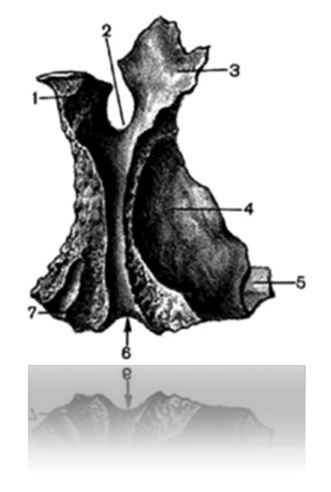
Fig. 24 Palatine bone (right) (external view)
641 214 918 516:
1 – sphenoid process 584 218 649 317
2 – sphenopalatine notch 549 641 219 811
3 – orbital process 549 674 317 581
4 – perpendicular plate 648 171 219 549
5 – horizontal plate 691 814 217 318
6 – greater palatine groove 394 698 598 714
7 – pyramidal process 691 218 514
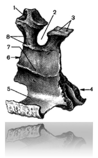
Fig. 25 Palatine bone, right (internal view)
649 314 219 516:
1 – orbital process 549 674 317 581
2 – sphenopalatine notch 549 612 814 914
3 – sphenoid process 584 218 649 317
4 – pyramidal process 691 218 514 317
5 – horizontal plate 691 814 217 318
6 – perpendicular plate 648 171 219 549
7 – concha comb 698 314 218 597
8 – lattice comb 467 214 909 814
Teeth 698 314 819 516
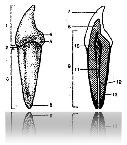
Fig. 26 General structure of the tooth:
1 – tooth crown 319 594 938 716
2 – tooth neck 364 891 219 491
3 – tooth root 368 549 188 794
4 – dental tubercle 364 198 501 248
5 – belt 601 549 906 714
6 – root apex of the tooth 594 315 498 515
7 – dental enamel 618 374 898 161
8 – dentin, dental substance 548 314 819 716
9 – dental pulp, dental pulp 316 481 219 649
10 – crown pulp 318 691 378 549
11 – root pulp 471 649 398 591
12 – root canal of the tooth 894 160 498 497
13 – cement 314 861 219 492
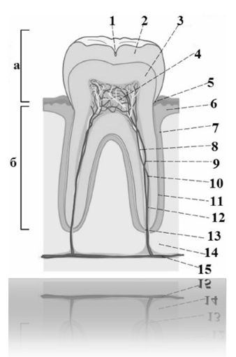
Fig. 27 Structure of the tooth and surrounding tissues:
a – crown 319 594 938 716
b - root 368 549 188 794
1 – fissure 546 218 319 491
2 – dental enamel 618 374 898 161
3 – dentin 548 314 819 716
4 – pulp 316 481 219 649
5 – gingival groove 518 316 549 471
6 – gums 479 168 318 517
7 – periodontium 482 316 219 491
8 – nerve fibers 478 514 219 671
9 – arterial vessels 894 378 214 316
10 – venous vessels 319 681 214 784
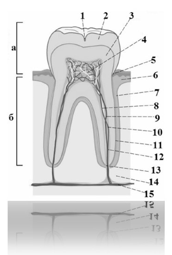
11 – root cement 698 541 349 172
12 – root canal of the tooth 894 160 498 497
13 – apical foramen 485 694 319 718
14 – jaw bone 318 549 468 019
15 – main vascular-nerve bundle
589 314 694 817
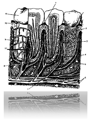
Fig. 28 Structure of the maxillofacial segment:
1 – gingival fibers 461 519 819 479
2 – alveolar wall 584 216 319 481
3 – dental-alveolar fibers 584 167 219 491
4 – alveolar-gingival branch 479 581 316 594
5 – periodontal vessels 891 478 219 641
6 – artery and veins of the jaw 984 517 219 648
7 – dental branch of the nerve794 281 298 641
8 – alveolar floor 514 368 791 498
9 – tooth root 368 549 188 794
10 – tooth neck 364 891 219 491
11 – tooth crown 319 594 938 716
12 – interdental (interroot) fibers
314 548 914 281
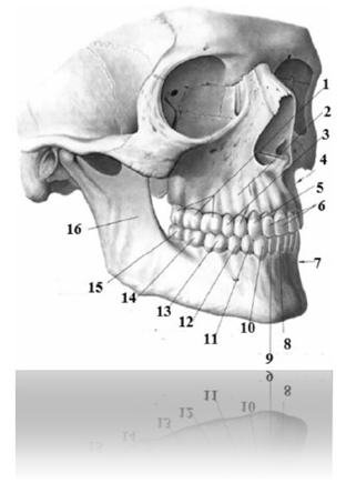
Fig. 29 Teeth of the upper and lower jaws, permanent
(right) 594 819 498 716:
1 – upper molars (from left to right) 648 517 216 318
- upper molar III 498 516 318 914
- upper molar II 548 491 478 694
- upper molar I 369 481 319 478
2 – alveolar elevations of the upper jaw
594 318 498 614
3 – upper premolars (from left to right)
694 317 219 498
- upper premolar II 378 498 514 916
- upper premolar I 614 218 598 781
4 – alveolar process of the upper jaw 986 149 318 518
5 – upper canine 471 891 016 498
6 – upper incisors (from left to right)519 671 918 549
-lateral incisor 549 691 718 548
- medial incisor 914 501 604 981
7 – alveolar part of the lower jaw 519 317 218 498
8 – lower medial incisor 584 716 914 219
9 – lower lateral incisor 989 718 514 601
10 – lower canine 589 318 499 164
11 – lower premolar I 518 016 949 148
12 – lower premolar II
514 817 316 498
13 – lower molar I 518 495 319 816
14 – lower molar II 519 814 317 984
15 – lower molar III 541 219 016 898
16 – lower jaw 514 712 814 312
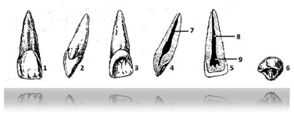
Fig. 30 Medial upper incisor (right)
914 501 604 981:
1 – vestibular surface 514 568 219 317
2 – mesial surface 314 894 319 892
3 – lingual surface 598 471 219 648
4 – internal view of the tooth in the vestibulo-lingual
planes 914 818 016 594
5 – internal view of the tooth in the mesio-distal
planes 317 648 519 819
6 – cutting surface 498 317 219 491
7 – root canal of the tooth 584 641 718 547
8 – root pulp 910 849 189 647
9 – crown pulp 319 648 519 987
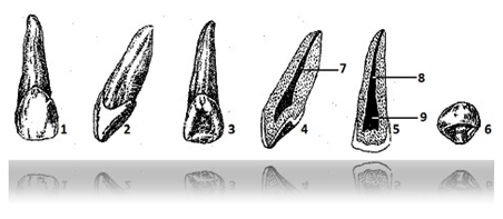
Fig. 31 Lateral upper incisor (right)
549 691 718 548:
1 – vestibular surface 514 916 917 518
2 – mesial surface 498 614 819 594
3 – lingual surface 718 594 319 681
4 – internal view of the tooth in the vestibulo-lingual
planes 594 716 918 916
5 – internal view of the tooth in the mesio-distal
planes 549 817 394 617
6 – cutting surface 581 349 619 817
7 – root canal of tooth 398 641 594 818
8 – root pulp 318 691 219 348
9 – crown pulp 819 601 698 149
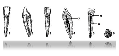
Fig. 32 Medial lower incisor (right)
584 716 914 219:
1 – vestibular surface 496 198 216 291
2 – mesial surface 481 478 594 316
3 – lingual surface 894 594 168 917
4 – internal view of the tooth in the vestibulo-lingual
planes 198 649 319 641
5 – internal view of the tooth in the mesio-distal
planes 894 167 318 491
6 – cutting surface 364 517 219 581
7 – root canal of tooth 318 694 369 471
8 – root pulp 949 516 817 919
9 – crown pulp 949 190 649 871
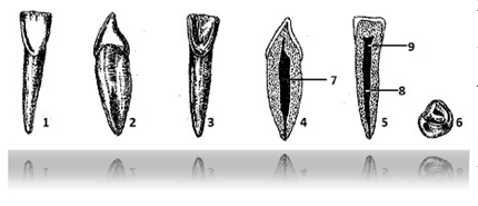
Fig. 33 Lateral lower incisor (right)
989 718 514 601:
1 – vestibular surface 694 187 219 471
2 – mesial surface 468 271 398 497
3 – lingual surface 894 561 219 718
4 – internal view of the tooth in the vestibulo-lingual
planes 584 617 219 714
5 – internal view of the tooth in the mesio-distal
planes 689 318 514 712
6 – cutting surface799 814 218 564
7 – root canal of tooth 368 194 371 894
8 – root pulp 964 718 519 498
9 – crown pulp 698 318 564 917
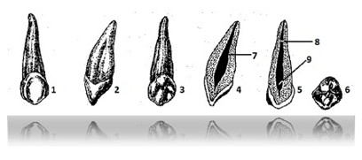
Fig. 34 Upper canine (right) 471 891 016 498:
1 – vestibular surface 468 716 519 498
2 – mesial surface 618 471 219 472
3 – lingual surface 549 316 218 581
4 – internal view of the tooth in the vestibulo-lingual
planes 918 516 319 491
5 – internal view of the tooth in the mesio-distal
planes 819 604 916 989
6 – cutting surface 641 519 318 491
7 – root canal of the tooth 384 591 689 374
8 – root pulp 989 618 054 132
9 – crown pulp 968 108 604 271
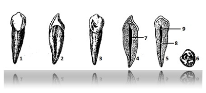
Fig. 35 Lower canine (right) 589 318 499 164:
1 – vestibular surface 698 318 514 217
2 – mesial surface 319 481 318 641
3 – lingual surface 948 564 008 904
4 – internal view of the tooth in the vestibulo-lingual
planes 368 014 218 548
5 – internal view of the tooth in the mesio-distal
planes 648 781 949 064
6 – cutting surface 546 981 941 568
7 – root canal of the tooth 714 801 498 541
8 – root pulp 398 614 718 581
9 – crown pulp 689 841 598 671
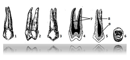
Fig. 36 First upper premolar (right)
614 218 598 781:
1 – vestibular surface 691 378 594 971
2 – mesial surface 498 617 898 541
3 – lingual surface 894 671 219 818
4 – internal view of the tooth in the vestibulo-lingual
planes 594 317 589 171
5 – internal view of the tooth in the mesio-distal
planes 478 641 219 891
6 – chewing surface 364 810 068 901
7 – root canals of the tooth 301 514 609 891
8 – root pulp 478 514 618 717
9 – crown pulp 984 018 198 601
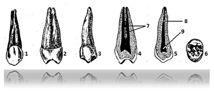
Fig. 37 Second upper premolar (right)
378 498 514 916:
1 – vestibular surface 948 561 319 818
2 – mesial surface 319 801 498 561
3 – lingual surface 467 219 498 541
4 – internal view of the tooth in the vestibulo-lingual
planes 398 548 589 617
5 – internal view of the tooth in the mesio-distal
planes 549 819 319 616
6 – chewing surface 389 541 379 818
7 – root canals of the tooth 648 546 319 818
8 – root pulp 894 361 219 012
9 – crown pulp 064 541 218 317
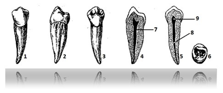
Fig. 38 First lower premolar (right)
518 016 949 148:
1 – vestibular surface 491 679 318 541
2 – mesial surface 549 361 819 497
3 – lingual surface 469 817 318 541
4 – internal view of the tooth in the vestibulo-lingual
planes 918 541 219 678
5 – internal view of the tooth in the mesio-distal
planes 814 319 898 514
6 – chewing surface 518 618 319 714
7 – root canal of the tooth 589 491 319 614
8 – root pulp 214 819 318 617
9 – crown pulp 914 218 519 641
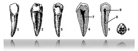
Fig. 39 Second lower premolar (right)
514 817 316 498:
1 – vestibular surface 698 517 319 641
2 – mesial surface 498 549 617 218
3 – lingual surface 894 317 218 491
4 – internal view of the tooth in the vestibulo-lingual
planes 469 518 519 641
5 – internal view of the tooth in the mesio-distal
planes 898 416 019 848
6 – chewing surface 841 319 718 491
7 – root canal of tooth 198 741 894 848
8 – root pulp 168 571 219 491
9 – crown pulp 371 549 619 814
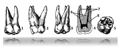
Fig. 40 First upper molar (right)
369 481 319 478:
1 – vestibular surface 491 614 718 541
2 – mesial surface 849 516 219 491
3 – lingual surface 684 517 919 486
4 – internal view of the tooth in the vestibulo-lingual
planes 584 619 319 814
5 – chewing surface 714 318 519 491
6 – root canals of the tooth 584 168 319 817
7 – root pulp 849 516 914 971
8 – crown pulp 318 619 819 498
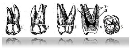
Fig. 41 Second upper molar (right)
548 491 478 694:
1 – vestibular surface 984 316 219 491
2 – mesial surface 894 518 319 491
3 – lingual surface 914 816 317 498
4 – internal view of the tooth in the vestibulo-lingual
planes 467 548 919 814
5 – chewing surface 214 391 898 491
6 – root canals of the tooth 316 598 368 498
7 – root pulp 648 718 598 647
8 – crown pulp 894 517 219 498
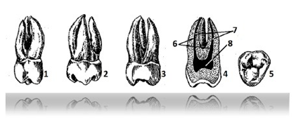
Fig. 42 Third upper molar (right)
498 516 318 914:
1 – vestibular surface 618 317 319 641
2 – mesial surface 689 064 194 818
3 – lingual surface 549 618 598 641
4 – internal view of the tooth in the vestibulo-lingual
planes 594 198 574 891
5 – chewing surface 648 591 318 498
6 – root canals of the tooth 491 684 898 718
7 – root pulp 964 717 988 149
8 – crown pulp 691 948 584 161
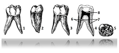
Fig. 43 First lower molar (right)
518 495 319 816:
1 – vestibular surface 319 681 519 894
2 – mesial surface 594 895 619 548
3 – lingual surface 694 171 218 541
4 – internal view of the tooth in the vestibulo-lingual
planes 549 614 318 541
5 – chewing surface 364 918 598 714
6 – root canals of the tooth 398 617 218 541
7 – root pulp 368 914 898 516
8 – crown pulp 319 891 498 516
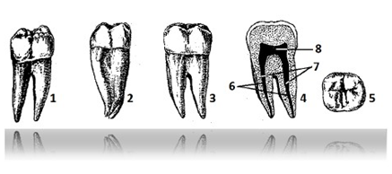
Fig. 44 Second lower molar (right)
519 814 317 984:
1 – vestibular surface 419 815 319 641
2 – mesial surface 498 316 318 541
3 – lingual surface 398 814 516 817
4 – internal view of the tooth in the vestibulo-lingual
planes 648 512 319 649
5 – chewing surface 504 194 981 369
6 – root canals of the tooth 894 016 598 641
7 – root pulp 897 491 219 896
8 – crown pulp 649 197 598 621
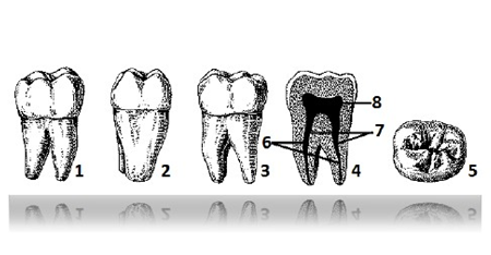
Fig. 45 Third lower molar (right)
541 219 016 898:
1 – vestibular surface 617 218 219 491
2 – mesial surface 694 817 219 497
3 – lingual surface 694 181 364 971
4 – internal view of the tooth in the vestibulo-lingual
planes 598 564 319 916
5 – chewing surface 948 516 218 949
6 – root canals of the tooth 319 491 819 647
7 – root pulp 384 161 219 491
8 – crown pulp 489 516 219 496
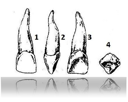
Fig. 46 Milk incisor, medial, upper
(right) 491 518 614 917:
1 – vestibular surface 549 618 219 814
2 – mesial surface 497 148 684 598
3 – lingual surface 248 379 064 898
4 – cutting surface 491 897 319 648
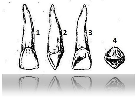
Fig. 47 Milk incisor, lateral, upper
(right) 514 218 919 648:
1 – vestibular surface 894 161 917 219
2 – mesial surface 619 517 319 498
3 – lingual surface 689 142 398 191
4 – cutting surface 218 589 649 171
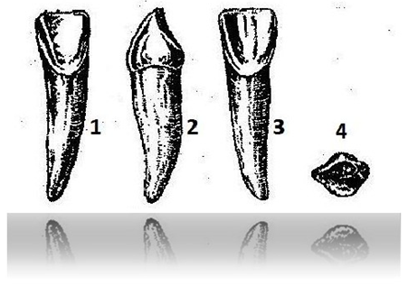
Fig. 48 Milk incisor, medial, lower
(right) 584 917 219 498:
1 – vestibular surface 514 817 219 648
2 – mesial surface 491 318 598 641
3 – lingual surface 461 598 597 681
4 – cutting surface 364 891 989 641
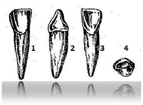
Fig. 49 Milk incisor, lateral,
lower (right) 549 817 219 491:
1 – vestibular surface 589 314 898 614
2 – mesial surface 386 149 948 511
3 – lingual surface 064 018 549 898
4 – cutting surface 414 818 619 710
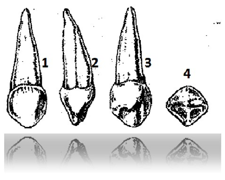
Fig. 50 Milk canine, upper (right)
498 691 798 541:
1 – vestibular surface 461 318 518 491
2 – mesial surface 498 641 319 814
3 – lingual surface 467 891 218 541
4 – cutting surface 318 491 819 617
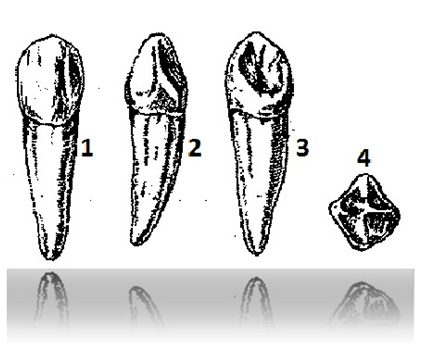
Fig. 51 Milk canine, lower (right)
619 317 218 491:
1 – vestibular surface 218 491 319 614
2 – mesial surface 214 817 218 316
3 – lingual surface 648 517 219 491
4 – cutting surface 314 817 219 617
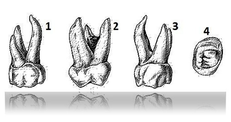
Fig. 52. First upper deciduous molar
(right):
1 – vestibular surface 491 718 519 497
2 – mesial surface 519 491 619 819
3 – lingual surface 594 817 219 648
4 – chewing surface 948 218 319 681
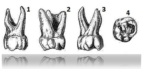
Fig. 53 Primary molar, second, upper
(right) 594 168 319 491:
1 – vestibular surface 619 518 219 491
2 – mesial surface 469 518 319 641
3 – lingual surface 584 316 589 491
4 – chewing surface 891 498 319 617
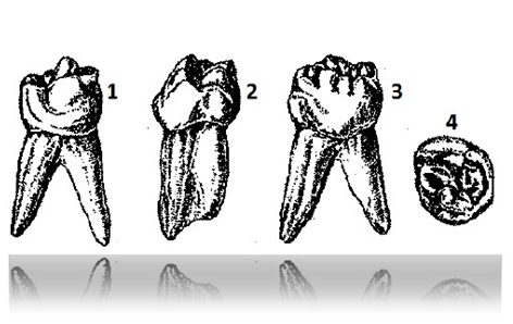
Fig. 54. First lower deciduous molar
(right) 491 318 519 491:
1 – vestibular surface 614 817 219 817
2 – mesial surface 486 519 719 491
3 – lingual surface 801 698 598 641
4 – chewing surface 894 219 319 810
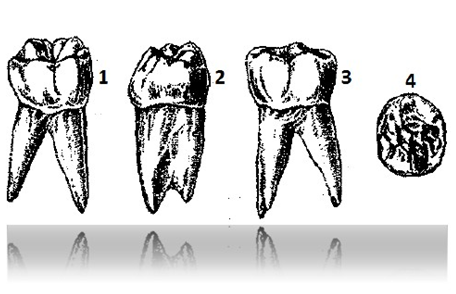
Fig. 55. Second lower deciduous molar
(right) 916 849 319 496:
1 – vestibular surface514 217 218 494
2 – mesial surface 584 564 819 718
3 – lingual surface 496 549 891 548
4 – chewing surface 649 817 918 491
Organs of the oral cavity
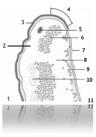
Fig. 56 Lip 498 718 494 814:
1 – inner side of the lip (oral cavity)
496 849 316 714
2 – submucosa 498 516 219 314
3 – mucous 589 641 218 549
4 – vermillion 598 714 219 674
5 – labial artery 598 541 219 491
6 – orbicularis oris muscle 548 321 818 221
7 – epidermis 598 718 889 888
8 – subcutaneous fat 594 817 549 164
9 – taste glands 198 016 219 491
10 – salivary glands 584 106 294 647
11 – outer side of the lip (skin) 319 891 498 647
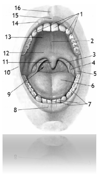
Fig. 57 Oral cavity and pharynx 489 461 319 891:
1 – upper dental arch 514 618 519 714
2 – palate suture 318 549 219 641
3 – palatopharyngeal arch 549 174 819 714
4 – palatine tonsil 514 218 319 671
5 – palatoglossus arch 479 604 594 219
6 – back of tongue 489 617 218 481
7 – lower dental arch 518 317 219 416
8 – lower lip 549 618 317 491
9 – pharynx 584 317 894 517
10 – lip commissure 584 316 318 497
11 – uvula (palatine) 314 841 219 647
12 – soft palate 549 561 718 649
13 – hard palate 564 817 219 481
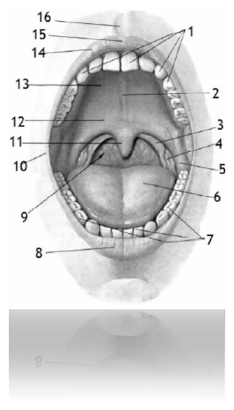
14 – upper lip 314 816 319 471
15 – tubercle of the upper lip 648 716 498 721
16 – Philtrum 642 148 894 216
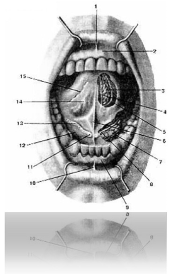
Fig. 58 Oral cavity 498 641 918 974
(Front view. Tongue raised. Material underneath.
mucous membrane):
1 – frenulum of the upper lip 498 691 719 497
2 – gum of the upper jaw 898 691 319 497
3 – anterior lingual gland 486 194 718 541
4 – lingual nerve 214 318 714 818
5 – inferior longitudinal muscle (of tongue) 319 648 319 781
6 – frenulum of the tongue 316 584 219 671
7 – sublingual gland 849 671 219 371
8 – submandibular duct 896 318 316 948
9 – gum of the lower jaw 519 318 219 641
10 – frenulum of the lower lip 318 364 891 871
11 – sublingual papilla 894 217 248 564
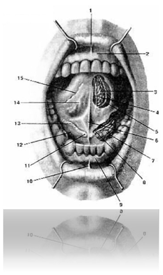
12 – floor (diaphragm) of the mouth 548 612 016 498
13 – sublingual fold 421 649 198 791
14 – lower surface of the tongue 219 064 284 714
15 – fringed fold 891 316 219 714
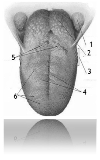
Fig. 59 Language 398 716 219 841:
1 – palatine tonsil 514 218 319 671
2 – lingual tonsil 498 791 648 219
3 – foliate papillae 418 644 319 515
4 – filiform papillae 589 617 298 471
5 – circumvallate papillae 891 319 481 617
6 – fungiform papillae 314 218 914 888
Chewing and facial muscles
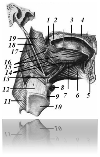
Fig. 60 Muscles of the tongue 594 218 598 641
(Right side view. Matter behind the right half
lower and upper jaws):
1 – palatoglossus muscle 194 891 319 491
2 – soft palate 549 561 718 649
3 – language 398 716 219 841
4 – hard palate 564 817 219 481
5 – lower jaw (part of the drawing) 514 712 814 312
6 – genioglossus muscle 218 614 319 718
7 – inferior longitudinal muscle (of tongue) 319 648 319 781
8 – hyoid bone 549 316 219 841
9 – median thyrohyoid ligament 598 617 219 641
10 – thyroid cartilage 549 891 364 218
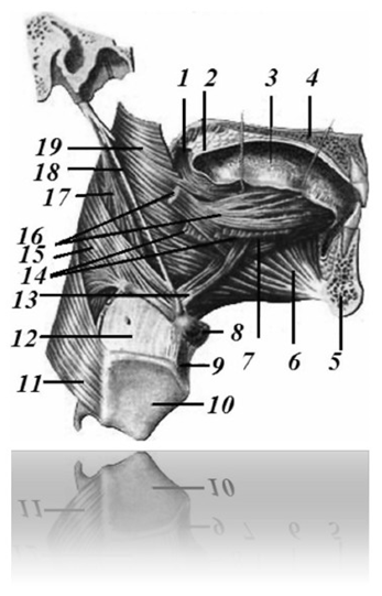
11 – inferior pharyngeal constrictor 584 216 234 271
12 – thyrohyoid membrane 584 691 219 478
13 – cartilaginous muscle 594 281 319 641
14 – hyoglossus muscle 814 316 498 384
15 – middle constrictor of the pharynx 548 314 894 851
16 – styloglossus muscle 584 391 314 891
17 – stylopharyngeus muscle 598 617 218 491
18 – stylohyoid ligament 584 217 278 061
19 – superior pharyngeal constrictor 348 541 618 714
Superficial facial muscles of the head 219 317 914 817
(part 1 fig. 102)
Facial muscles (front view) 598 641 398 719
(part 1 fig. 103)
Deep facial muscles 328 721 428 919
(part 1 fig. 104)
Masticatory muscles 519 314 819 214
(part 1 fig. 105)
Temporomandibular joint
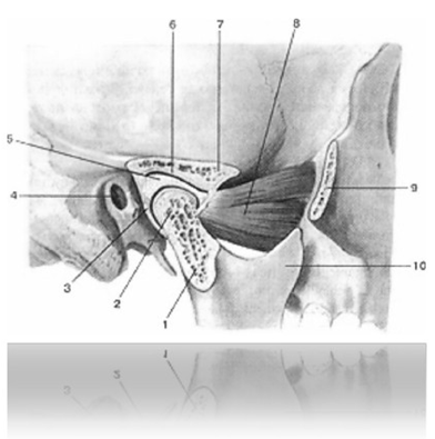
Fig. 61 Temporomandibular joint
(sagittal projection):
1 – articular (condylar) process of the lower
jaws 891 319 898 789
2 – head of the lower jaw 548 321 848 721
3 – joint capsule 498 641 718 491
4 – external auditory canal 519 421 919 811
5 – articular (intra-articular) disc 894 516 219 497
6 – mandibular fossa 558 912 918 222
7 – articular tubercle 288 412 298 322
8 – lateral pterygoid muscle 219 214 319 214
9 – temporal process of the zygomatic bone 694 171 219 548
10 – coronoid process of the lower jaw
528 317 918 228
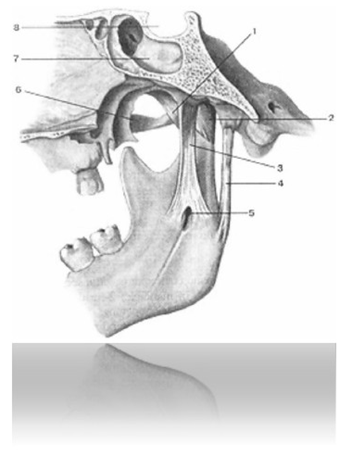
Fig. 62 Ligaments of the temporomandibular joint
819 491 319 848 (medial view):
1 - lateral ligament (temporomandibular
joint) 519 647 218 541
2 – capsule of the temporomandibular joint
498 641 718 491
3 – sphenomandibular ligament 584 317 219 497
4 – awl-mandibular ligament 898 514 518 316
5 – opening of the lower jaw 489 201 319 871
6 – zygomatic arch 528 317 918 917
7 – sphenoid sinus 584 217 319 841
8 – pituitary fossa (sella turcica) 519 317 919 218
Glands of the vestibule and oral cavity 498 617 219 491
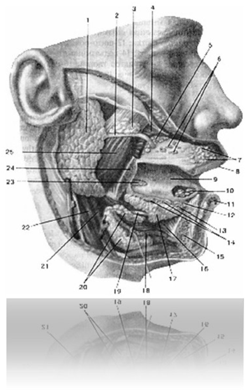
Fig. 63 Glands of the vestibule and oral cavity
498 617 219 491 (right view):
1 – parotid gland 194 817 219 418
2 – parotid duct 218 491 619 317
3 – accessory parotid gland 514 816 719 497
4 – cheek muscle 549 317 849 217
5 – molar glands 514 817 219 498
6 – cheek glands 548 742 819 461
7 – labial glands 548 649 319 817
8 – upper lip 314 816 319 471
9 – language 398 716 219 841
10 – anterior lingual gland 486 194 718 541
11 – lower lip 549 618 317 491
12 – sublingual papilla 894 316 598 718
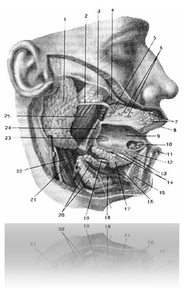
13 – greater sublingual duct 548 717 219 418
14 – small sublingual ducts 498 641 318 374
15 – lower jaw 514 712 814 312
16 – genioglossus muscle 218 614 319 718
17 – sublingual gland 849 671 219 371
18 – mylohyoid muscle 498 541 316 841
19 – submandibular duct 896 318 316 948
20 – submandibular gland 498 714 319 481
21 – stylohyoid muscle 594 217 298 647
22 – posterior belly of the digastric muscle
316 849 918 716
23 – posterior lingual gland 314 849 216 371
24 – lower jaw 514 712 814 312
25 – chewing muscle 598 712 918 212
SPINE.
JOINTS, LIGAMENTS AND MUSCLES OF THE SPINE
Vertebral column 214 217 000 819
(continued) (part 1 fig. 62)
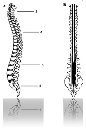
Fig. 64 Vertebral column
214 217 000 819 (continued):
A - Physiological curves of the spine
598 614 818 017
Primary bends 319 892 964 718
2 – thoracic kyphosis 379 491 814 219
4 – sacral kyphosis 598 061 719 898
Secondary bends 598 718 419 061
1 – cervical lordosis 898 716 919 041
4 – lumbar lordosis 584 061 718 910
B – Spinal canal 521 314 818 214
VERTEBRAE 498 641 319 048
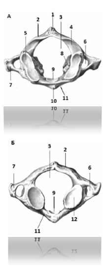
Fig. 65 First cervical vertebra (atlas)
914 816 978 496:
A - top view
B - bottom view
1 – posterior tubercle 894 217 319 498
2 – rear arch 894 617 319 497
3 – vertebral foramen 864 914 898 516
4 – groove of the vertebral artery 749 891 218 641
5 – superior glenoid fossa 598 691 219 674
6 – transverse hole (cross hole)
process) 649 581 219 697
7 – transverse process 584 316 918 581
8 – lateral mass 648 719 218 541
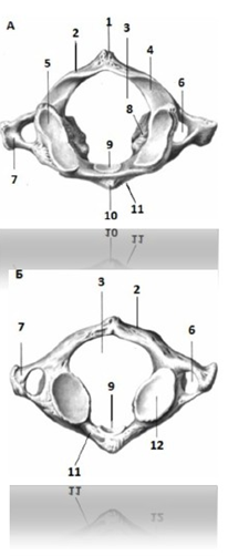
9 – tooth pit 694 197 289 471
10 – anterior tubercle 319 691 218 712
11 – front arch 649 171 218 641
12 – inferior glenoid fossa 598 317 294 817
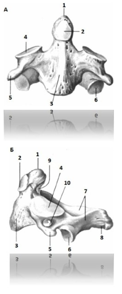
Fig. 66 Second cervical vertebra (axial)
794 218 849 617:
A - front view
B - side view
1 – tooth of the axial vertebra 598 314 219 617
2 – anterior articular surface 698 591 219 491
3 – vertebral body 598 674 218 514
4 – superior articular process 589 491 218 641
5 – transverse process 698 371 294 811
6 – inferior articular process 541 319 894 361
7 – vertebral arch 898 561 219 364
8 – spinous process 581 319 619 714
9 – posterior articular surface 598 612 819 498
10 – transverse hole (cross hole)
process) 594 612 898 714
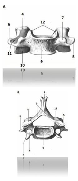
Fig. 67 Cervical vertebra (III – VI cervical
vertebrae) 498 317 218 641:
A - front view
B - top view
1 – spinous process 514 217 218 684
2 – vertebral foramen 319 648 281 317
3 – vertebral arch 094 701 278 649
4 – superior articular process 894 361 219 897
5 – transverse process 698 317 298 641
6 – posterior tubercle of the transverse process
550 694 931 074
7 – anterior tubercle of the transverse process
894 171 219 647
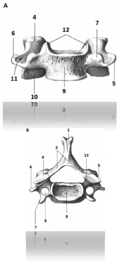
8 – transverse hole (cross hole)
process) 589 316 298 649
9 – vertebral body 368 174 289 691
10 – inferior articular process 519 581 314 891
11 – groove of spinal nerve 649 718 219 417
12 – body hook 689 517 219 618
13 – superior vertebral notch 216 541 319 714
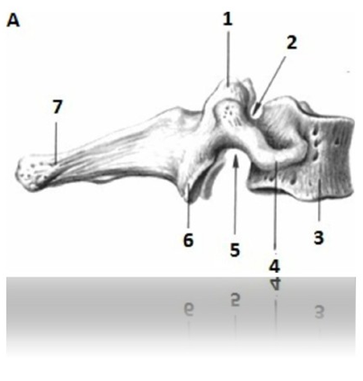
Fig. 68 Seventh cervical vertebra:
319 648 519 647:
A - side view
B - top view
1 – superior articular process 584 216 549 617
2 – superior vertebral notch 594 691 798 714
3 – vertebral body 918 694 319 896
4 – transverse process 698 712 319 641
5 – lower vertebral notch 549 598 694 714
6 – inferior articular process 548 217 219 691
7 – spinous process 591 316 214 278
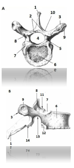
Fig. 69 Thoracic vertebra 542 317 212 227:
A - top view
B - side view
1 – spinous process 518 617 218 141
2 – vertebral arch 648 549 819 712
3 – transverse process 598 642 319 811
4 – vertebral foramen798 621 319 416
5 – pedicle of the vertebral arch 498 317 218 217
6 – vertebral body 517 219 319 617
7 – superior costal fossa 549 312 814 212
8 – superior articular process 219 715 319 215
9 – transverse costal fossa (costal fossa
transverse process) 821 319 921 819
10 – vertebral arch plate 514 218 619 719
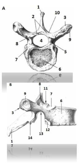
11 – superior vertebral notch 598 641 398 011
12 – lower costal fossa 019 712 219 312
13 – lower vertebral notch 512 314 812 214
14 – inferior articular process 528 644 328 016
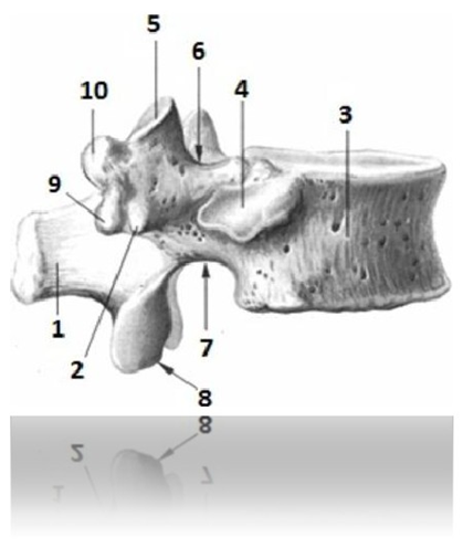
Fig. 70 XII Thoracic vertebra (XI and XII thoracic
vertebrae) (side view) 496 819 318 514:
1 – spinous process 314 815 619 718
2 – transverse process 218 316 514 471
3 – vertebral body 364 819 519 614
4 – costal fossa 818 542 617 218
5 – superior articular process 514 618 019 008
6 – superior vertebral notch 194 691 298 511
7 – lower vertebral notch 584 317 218 584
8 – inferior articular process 549 613 219 814
9 – additional process 319 684 218 514
10 – mastoid process 819 617 218 419
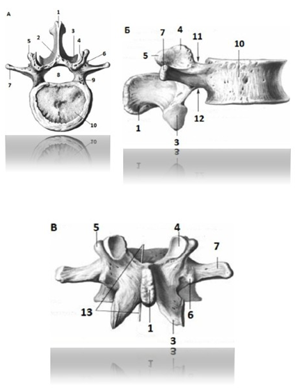
Fig. 71 Lumbar vertebra 618 513 219 418:
A - top view
B - side view
B - rear view
1 – spinous process 513 219 813 919
2 – vertebral arch 391 498 016 217
3 – inferior articular process 549 316 218 494
4 – superior articular process 519 617 299 017
5 – mastoid process 918 217 319 817
6 – additional process 518 431 219 917
7 – costal process 317 814 214 917
8 – vertebral foramen 828 317 918 217
9 – pedicle of the vertebral arch 498 317 218 217
10 – vertebral body 598 641 319 071
11 – superior vertebral notch 518 491 316 498
12 – lower vertebral notch 549 617 219 811
13 – vertebral foramen (projection of the foramen)
828 317 918 217
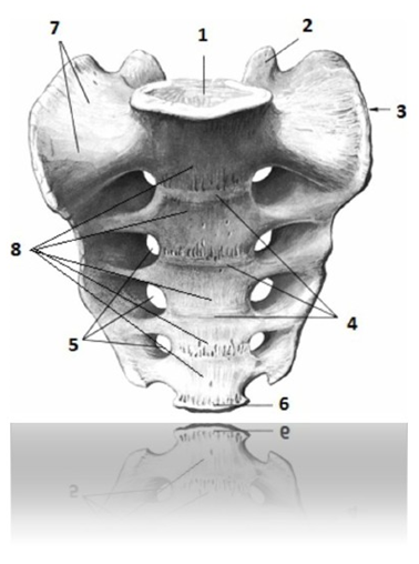
Fig. 72 Sacrum 514 716 814 226
(front view, pelvic surface):
1 – base of the sacrum 519 614 319 812
2 – superior articular process 519 328 919 228
3 – lateral part 319 712 919 212
4 – transverse lines 428 213 328 333
5 – anterior sacral openings 489 213 217 289
6 – apex of the sacrum 408 217 229 327
7 – sacral wing 519 618 514 217
8 – sacral vertebrae 584 317 218 498
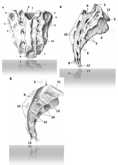
Fig. 73 Sacrum 514 716 814 226:
A – posterior view (dorsal surface)
B - side view
B - internal view in the median-longitudinal direction
planes
1 – sacral canal (upper opening)
594 647 289 391
2 – superior articular process 519 328 919 228
3 – sacral tuberosity 498 316 219 471
4 – ear-shaped surface 594 561 378 541
5 – lateral sacral crest 584 816 219 471
6 – intermediate sacral crest
319 641 281 491
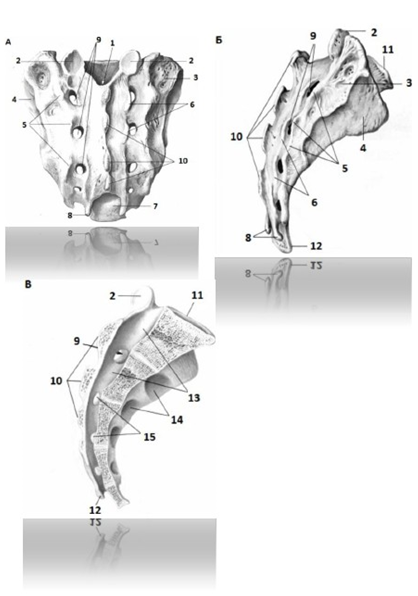
7 – sacral hiatus (lower opening of the sacral
channel) 316 218 319 091
8 – sacral horns 019 001 849 471
9 – dorsal (posterior) sacral openings
698 041 278 914
10 – median sacral crest 518 691 298 741
11 – base of the sacrum 519 614 319 812
12 – apex of the sacrum 408 217 229 327
13 – sacral canal 584 621 319 647
14 – anterior sacral openings 489 213 217 289
15 – intervertebral openings584 101 294 988
TAZ 584 316 719 041
Pelvic bone (right) (lateral view)
214 317 918 227 (part 1, fig. 89A)
Pelvic bone (right) (medial view)
214 317 918 227 (part 1, fig. 89B)
Coccyx 519 513 819 213 (part 1, fig. 59)
Vertebral motor segment 714 986 219 694
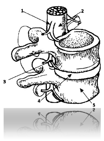
Fig. 74 Vertebral motor segment
714 986 219 694:
1 – nerve root 519 691 219 814
2 – spinal cord 314 218 814 719
3 – intervertebral foramen 517 218 916 284
4 – intervertebral disc 648 217 398 491
5 – vertebral body 849 161 219 711
Muscles and ligaments of the spine 549 641 894 217
Spinal column joints 894 216 819 048
Syndesmoses (ligaments) of the spinal column 398 947 019 818
Synchondrosis of the spinal column 519 312 498 061
Joints of the spine 719 891 498 061
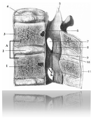
Fig. 75 Vertebral joints
(sagittal projection at the level of two
lumbar vertebrae) 498 641 917 218:
A – intervertebral symphysis 898 064 317 219
B – facet joint 598 071 319 481
1 – vertebral body 598 641 319 071
2 – gelatinous nucleus of the intervertebral disc 514 891 518 316
3 – anterior longitudinal ligament 689 174 219 814
4 – fibrous ring of the intervertebral disc 498 716 219 714
5 – superior articular process of the lumbar vertebra
519 617 299 017
6 – posterior longitudinal ligament 548 691 218 781
7 – intervertebral foramen of the lumbar region
spine 916 048 219 491
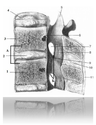
8 – yellow ligament 549 488 194 016
9 – articular capsule of the facet joint (intervertebral)
joint 364 198 278 471
10 – interspinous ligament 368 142 894 216
11 – supraspinous ligament 890 149 540 691
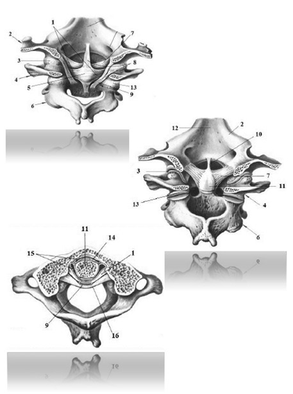
Fig. 76 Joints between the occipital bone and
I-II cervical vertebrae 379 814 919 718:
1 - transverse ligament of the first cervical vertebra
589 061 319 498
2 – occipital bone 214 712 219 312
3 – atlanto-occipital joint 591 048 319 491
4 – I cervical vertebra 914 816 978 496
5 – cruciate ligament of the atlas 618 717 919 064
6 – II cervical vertebra 794 218 849 617
7 – alar ligament 598 019 318 941
8 – longitudinal beams 541 061 719 801
9 – Covering membrane 391 848 319 064
10 – ligament of the apex of the tooth 718 391 898 491
11 – tooth of the axial vertebra 598 314 219 617
12 – slope of the base of the skull 319 778 219 228
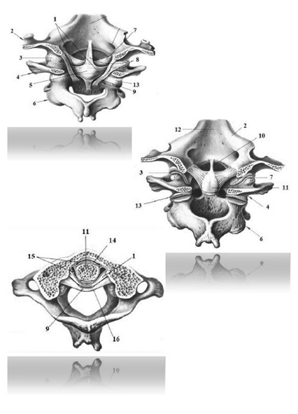
13 – lateral atlantoaxial joint
719 891 906 217
14 – median atlantoaxial joint
598 089 319 641
15 – articular cavity of the median atlantoaxial
joint 489 061 918 217
16 – posterior longitudinal ligament 384 619 818 061
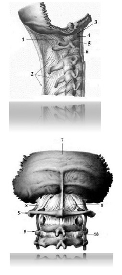
Fig. 77 Ligaments of the cervical vertebrae and occipital bone
718 119 498 064:
1 – posterior atlanto-occipital membrane 598 817 319 048
2 – nuchal ligament 517 319 049 811
3 – anterior atlanto-occipital ligament 490 391 849 061
4 – anterior atlanto-occipital membrane 598 601 819 317
5 – I cervical vertebra 914 816 978 496
6 – lateral atlantoaxial joint 719 891 906 217
7 – occipital bone 214 712 219 312
8 – lateral atlanto-occipital ligament 974 217 298 041
9 – II cervical vertebra 794 218 849 617
10 – yellow ligament 549 488 194 016
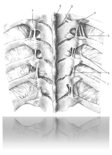
Fig. 78 Ligaments of the spine (thoracic region) and
costovertebral joints
514 891 219 478:
1 – ligament of the costal tubercle 698 714 219 811
2 – supraspinous ligament 890 149 540 691
3 – yellow ligament 549 488 194 016
4 – costotransverse ligament 948 691 219 794
5 – lateral costotransverse ligament
894 691 217 474
6 – intertransverse ligaments 514 692 899 714
7 – internal intercostal membrane
598 726 319 491
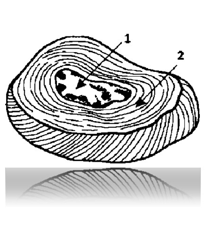
Fig. 79 Intervertebral disc 648 217 398 491:
1 – nucleus pulposus (gelatinous) 514 891 518 316
2 – fibrous ring 498 716 219 714
LIGAMENTS OF THE PELVIS AND HIP JOINT 498 641 798 478
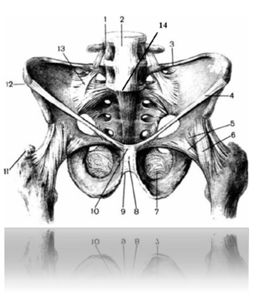
Fig. 80 Ligaments of the pelvis and hip joint
498 641 798 478:
A - front view
1 – IV lumbar vertebra 467 198 219 481
2 – anterior longitudinal ligament 848 471 219 819
3 – iliopsoas ligament 319 641 289 798
4 – inguinal ligament949 641 289 541
5 – joint capsule of the hip joint
589 671 218 498
6 – iliofemoral ligament 364 911 894 564
7 – locking membrane 312 689 319 716
8 – pubic symphysis 368 214 598 471
9 – arcuate pubic ligament 496 549 718 614
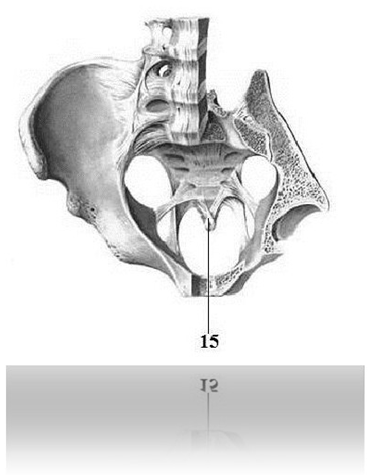
10 – superior pubic ligament 894 216 218 498
11 – Large spit 519 814 089 319
12 – anterior superior iliac spine
379 041 298 517
13 – ventral sacroiliac ligament
589 491 291 478
14 – lumbosacral joint 591 071 298 498
15 – anterior sacrococcygeal ligament
578 601 949 011
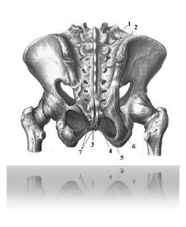
Fig. 80 Ligaments of the pelvis and hip joint
498 641 798 478:
B - rear view
1 – iliopsoas ligament 479 681 598 718
2 – dorsal sacroiliac ligament
574 981 319 818
3 - dorsal superficial sacrococcygeal
bundle 498 688 715 301
4 - dorsal deep sacrococcygeal
bundle 594 072 319 401
5 - dorsal lateral sacrococcygeal
bundle 719 317 908 481
6 – sacrotuberous ligament 501 489 714 211
7 – sacrococcygeal joint 291 081 407 201
MUSCLES AND FASCIA OF THE BACK AND OCCIUM 798 041 261 509
Superficial muscles of the back 819 314 914 812
(part 1 fig. 97)
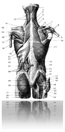
Fig. 81 Muscles of the back and posterior neck region (superficial
muscles, first, second and third layers) 519 648 218 741:
1 – semispinalis capitis muscle 914 217 218 498
2 – splenius capitis muscle 298 742 279 488
3 – splenius muscle of the neck 216 498 948 741
4 – muscle that lifts the scapula 214 317 914 717
5 – rhomboid minor muscle 319 061 919 618
6 – large rhomboid muscle 584 317 914 016
7 – supraspinatus muscle 312 214 812 514
8 – infraspinatus muscle (partial projection) 894 314 818 574
9 – small round muscle (part of the figure) 498 518 491 748
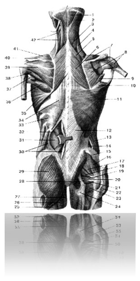
10 – large round muscle (part of the figure) 849 516 319 478
11 – latissimus dorsi; 429 318 829 998
12 – aponeurosis of the latissimus dorsi muscle; 549 718 219 478
13 – external oblique abdominal muscle 529 312 419 272
14 – lumbar triangle 894 568 514 811
15 – gluteal fascia 598 314 698 718
16 – gluteus medius 589 491 219 641
17 – gluteus minimus 364 917 584 218
18 – piriformis muscle 498 571 218 498
19 – superior gemellus muscle 364 581 219 644
20 – internal obturator muscle 398 711 264 814
21 – inferior gemellus muscle 314 894 219 471
22 – gluteus maximus (part of the figure) 548 361 894 317
23 – quadratus femoris 694 584 219 471
24 – ischial tuberosity 529 312 918 812
25 – biceps femoris 598 617 329817
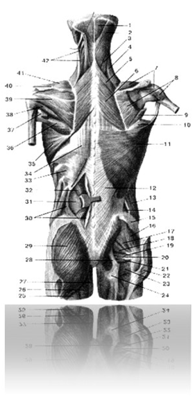
26 – semitendinosus muscle 549 381 714 817
27 – adductor magnus 374 841 219 471
28 – sacrotuberous ligament 316 497 218 914
30 – superficial leaflet of the fudolumbar fascia (projection
with a side flap) 494 848 514 216
31 – deep layer of the thoracolumbar fascia 481 319 614 714
32 – muscle that straightens the trunk (pulled to the medial
side) 598 748 519 491
33 – inferior posterior serratus muscle 549 317 919 817
34 – fudo-lumbar fascia 529 317 919 817
35 – serratus anterior muscle 219 475 819 355
36 – large round muscle 849 516 319 478
37 – long head of the triceps brachii (part of the figure)
514 819 498 614
38 – small round muscle 498 518 491 748
39 – infraspinatus muscle 894 314 818 574
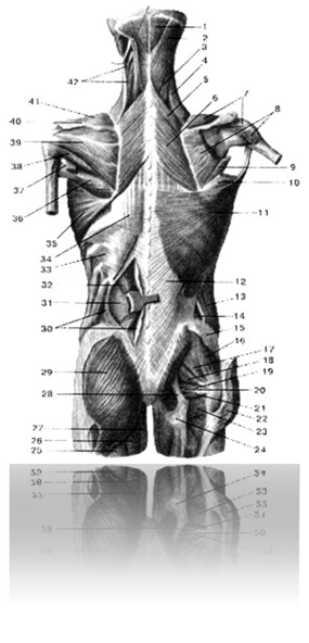
40 – scapula spine 498 712 328 822
41 – supraspinatus muscle 312 214 812 514
42 – muscle that lifts the scapula (pulled to the side)
214 317 914 717
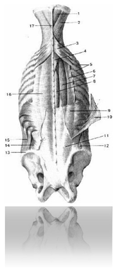
Fig. 82 Muscles of the back and neck 498 549 618 714
(the muscles and bones of the shoulder girdle are not shown in the picture
shown):
1 – semispinalis capitis muscle 914 217 218 498
2 – splenius capitis muscle 298 742 279 488
3 – superior posterior serratus muscle 898 549 694 714
4 – splenius muscle of the neck 216 498 948 741
5 – external intercostal muscles 398 591 294 168
6 – iliocostalis dorsi muscle 319 647 218 471
7 – longest back muscle 497 549 819 714
8 – spinal muscle 396 891 319 471
9 – lower posterior serratus muscle 549 317 919 817
10 – latissimus dorsi (part of the drawing, turned away)
side) 429 318 829 998
11 – aponeurosis of the latissimus dorsi muscle 549 718 219 478
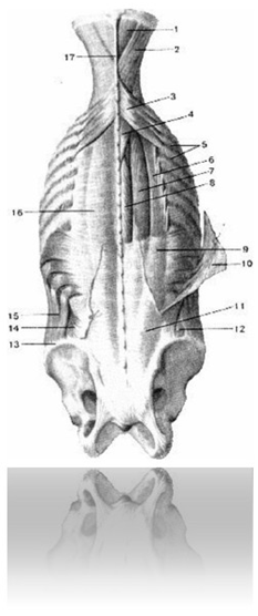
12 – lumbar triangle 894 568 514 811
13 – iliac crest 894 547 218 471
14 – internal oblique muscle of the abdomen 398 217 818 417
15 – external oblique abdominal muscle 529 312 419 272
16 – thoracolumbar fascia 584 317 019 641
17 – nuchal ligament 589 691 319 714
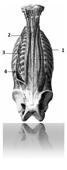
Fig. 83 Iliocostalis muscle 314 841 619 714:
1 – iliocostalis muscle 314 841 619 714
2 – iliocostalis muscle of the neck 491 481 471 819
3 – iliocostalis back muscle 584 461 489 714
4 – iliocostalis lumborum muscle 578 714 218 417
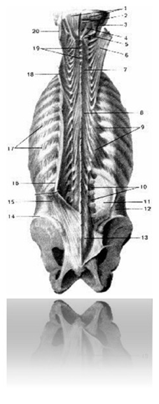
Fig. 84 Deep muscles of the back and neck 498 714 219 614:
1 – semispinalis capitis muscles 914 217 218 498
2 – rectus capitis posterior minor 219 817 819 227
3 – superior oblique muscle of the head 218 417 918 817
4 – large posterior rectus capitis muscle 594 318 614 715
5 – inferior oblique muscle of the head 218 317 918 227
6 – semispinalis capitis muscle (part of the picture, turned away)
914 217 218 498
7 – semispinalis cervicis muscle 319 714 218 412
8 – semispinalis muscle of the back 318 694 218 421
9 – muscles that raise the ribs 689 714 298 514
10 – intertransverse muscles 519 314 819 312
11 – deep layer of the thoracolumbar fascia 481 319 614 714
12 – transverse abdominal muscle 555 813 915 513
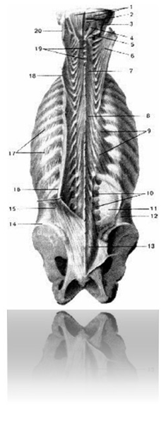
13 – multifidus muscle 549 781 219 471
14 – iliac wing 529 301 229 721
15 – iliocostalis muscle 364 712 819 418
16 – longissimus muscle 589 641 289 714
17 – external intercostal muscles 369 581 298 471
18 – longest muscle of the neck 699 186 019 491
19 – interspinous muscles of the neck 489 617 819 398
20 – longissimus capitis 389 497 368 141
Female pelvis 494 714 516 841
FEMALE GENITAL ORGANS 519 814 089 319
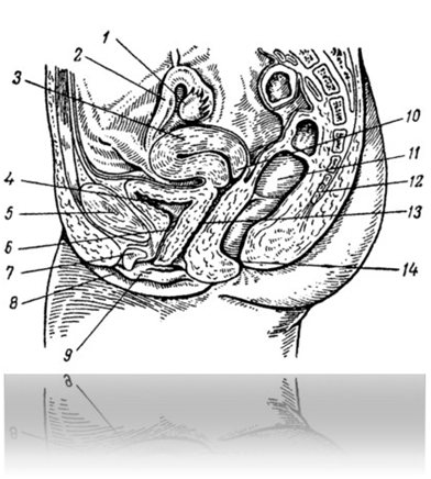
Fig. 85 Female genitalia 519 814 089 319
(longitudinal median projection):
1 – ovary 914 814 917 218
2 – fallopian tube 619 718 316 214
3 – body of the uterus 689 514 218 471
4 – urinary bladder 219 389 998 419
5 – pelvic bone (part of the drawing) 214 317 918 227
6 – urethra 329 487 948 216
7 – clitoris 689 568 319 818
8 – labia majora 598 711 008 512
9 – labia minora 319 016 789 498
10 – cervix 894 581 948 164
11 – rectum 598 714 898 314
12 – sacrum 514 716 814 226
13 – vagina 889 491 619 819
14 – anus 589 317 418 917
Female external genitalia 519 319 818 678
(part 1, fig 124)
Internal female genital organs 419 219 808 319
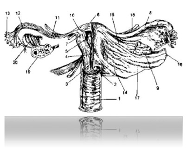
Fig. 86 Internal female genital organs
419 219 808 319:
1 – vagina 889 491 619 819
2 – vaginal part of the cervix 548 988 581 497
3 – cervical canal 614 891 719 489
4 – isthmus 549 691 289 784
5 – uterine cavity 318 688 594 191
6 – fundus of the uterus 984 016 501 348
7 – uterine wall 841 369519 471
8 – fallopian tube 619 718 316 214
9 – ovary 914 814 917 218
10 – interstitial part of the tube 584 199 598 641
11 – isthmic part of the pipe 589 612 319 471
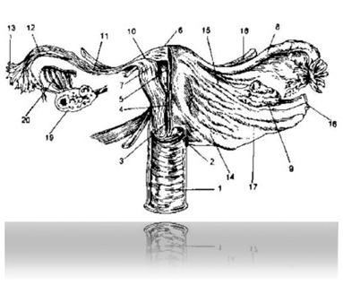
12 – ampullar part of the pipe 894 316 498 561
13 – fimbriae of the tube 589 617 289 748
14 – uterosacral ligament 598 361 298 471
15 – proper ovarian ligament 584 216 298 497
16 – infundibulopelvic ligament 294 147 284 641
17 – wide bundle 549 581 369 471
18 – round ligament 948 371 296 497
19 – projection of the ovary with follicles and corpus luteum
498 316 478 471
20 – steam room 891 368 194 364
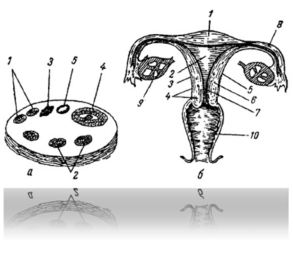
Fig. 87 Schematic representation of the longitudinal
projections of the ovary (a) and transverse
projections of the uterus (b):
A:
1 – primary follicle 498 518 818 491
2 – growing follicle 898 648 218 471
3 – white body 497 614 201 498
4 – Graafian bubble 481 684 371 016
5 – corpus luteum 818 401 616 214
b:
1 – fundus of the uterus 984 016 501 348
2 – uterine cavity 318 688 594 191
3 – body of the uterus 689 514 218 471
4 – cervix 894 581 948 164
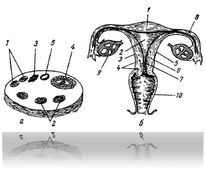
5 – outer layer of the uterus, serous membrane
(perimetry) 848 147 218 417
6 – middle layer of the uterus, muscular layer
(myometrium) 198 316 949 101
7 – the inner layer of the uterus, the mucous membrane
(endometrium) 698 317 281 488
8 – fallopian tube 619 718 316 214
9 – ovary 914 814 917 218
10 – vagina 889 491 619 819
MAMMARY GLAND 648317219491
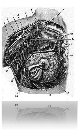
Fig. 88 Structure and topography of the mammary gland:
1 - lateral axillary lymph node
497 218 217 498
2 – axillary artery 694 718 217 491
3 – axillary vein 849 716 218 471
4 – brachial plexus 312 314 512 214
5 – central axillary lymph node
648 516 201 505
6 – apical axillary lymph nodes
618 471 298 741
7 – supraclavicular lymph nodes 598 641 264 271
8a – lateral thoracic artery 598 722 918 213
8b – lateral thoracic vein 584 317 248 517
9 – sternal lymph nodes 649 318 714 618
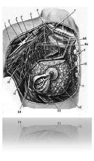
10 – plexus of blood and lymphatic vessels
510 314 784 617
11 – branch of the internal thoracic artery to the mammary gland
479 841 589 641
12 – areola 394 647 198 518
13 – milk ducts 471 691 284 714
14 – lateral arterial branches of the mammary gland
691 014 398 517
15 – thoracic axillary lymph nodes
749 148 519 618
16 – subscapular lymph nodes 184 816 014 214
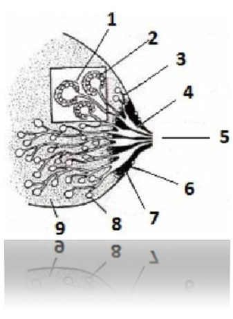
Fig. 89 Anatomy of the mammary glands:
1 – muscle cells 494 816 319 481
2 – milk-secreting cells 859 169 794 217
3 – milk ducts 471 691 284 714
4 – lactiferous sinuses 584 316 219 478
5 – nipple 894 181 319 718
6 – areola 394 647 198 518
7 – Montgomery glands 491 819 488 514
8 – alveoli 614 819 319 714
9 – supporting and adipose tissue 898 617 219 419
CARDIOVASCULAR SYSTEM
(continued) 214 700 819 891
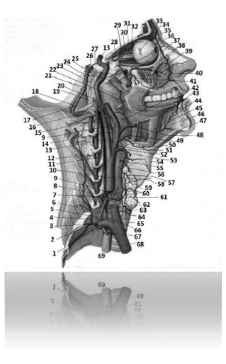
Fig. 90 Arteries of the head and neck (right view)
(continued) 518 422 819 312:
1 – subclavian artery (right) 598 317 819 227
2 – costocervical trunk 549 641 898 714
3 – highest intercostal artery 589 716 549 818
4 – thyrocervical trunk 519 317 919 288
5 – suprascapular artery 529 317 419 817
6 – deep cervical artery 598 714 898 716
7 – ascending cervical artery 749 814 719 414
8 – VI – cervical vertebra 568 314 819 714
9 – pharyngeal branches 498 217 228 417
10 – common carotid artery (right) 919 421 818 728
11 – vertebral artery (cervical part) 498 714 319 716
12 – spinal branches 584 314 819 417
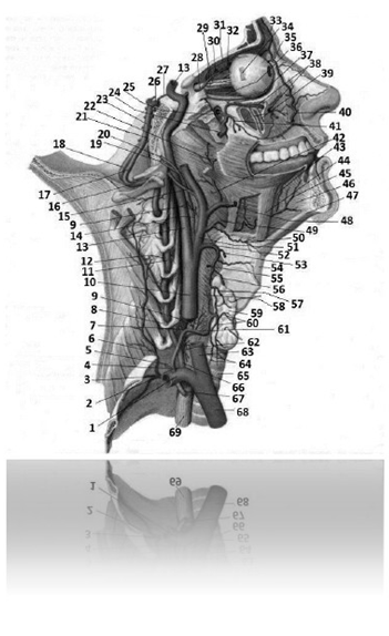
13 – internal carotid artery 549 712 810 248
14 – ascending pharyngeal artery 496 598 317 641
15 – occipital artery 581 214 608 491
16 – vertebral artery (cervico-occipital part)
918 317 948 561
17 – vertebral artery (right) (intracranial part)
364 819 498 471
18 – vertebral artery (left) (intracranial part)
364 819 498 471
19 – inferior tympanic artery 894 168 941 987
20 – posterior meningeal artery 594 162 398 714
21 – slope of the base of the skull 319 778 219 228
22 – amygdaloid branch 895 316 498 471
23 – basilar artery 851 478 594 814
24 – maxillary artery 648 517 284 917
25 – sphenopalatine artery 598 491 374 816
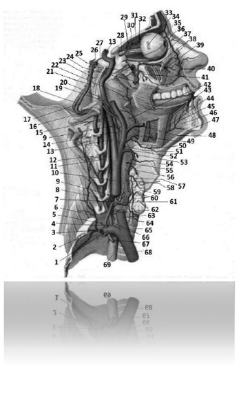
26 – posterior cerebral artery 594 361 809 491
27 – posterior communicating artery 548 641 298 781
28 – ophthalmic artery 649 718 549 641
29 – posterior short ciliary arteries 496 391 898 671
30 – posterior ethmoid artery 594 716 298 491
31 – supraorbital artery 594 617 548 518
32 – anterior ethmoid artery 698 713 294 168
33 – supratrochlear artery 798 791 694 814
34 – lateral rectus muscle of the eye 328 421 898 712
35 – dorsal nasal artery 368 142 598 714
36 – posterior long ciliary arteries 581 641 294 818
37 – inferior oblique muscle of the eye 319 618 204 881
38 – infraorbital artery 898 048 319 061
39 – anterior superior cellular arteries 364 181 298 471
40 – angular artery 288 919 069 789
41 – posterior superior cellular artery 549 161 298 191
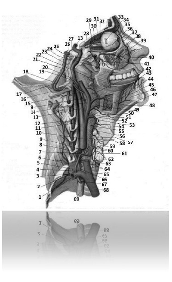
42 – ascending palatine artery 581 494 549 618
43 – deep artery of the tongue 319 694 384 716
44 – hyoglossus muscle 368 142 498 641
45 – sublingual artery 784 981 294 671
46 – genioglossus muscle 218 614 319 718
47 – Geniohyoid muscle 498 694 819 671
48 – facial artery 219 061 234 890
49 – lingual artery 498 519 401 209
50 – suprahyoid branch 691 318 464 148
51– external carotid artery 510 469 148 712
52 – superior thyroid artery 519 513 719 313
53 – superior laryngeal artery 389 461 894 171
54 – thyrohyoid membrane 584 691 219 478
55 – sternocleidomastoid branch 519 614 431 548
56 – anterior branch of the superior thyroid artery
698 517 401 469
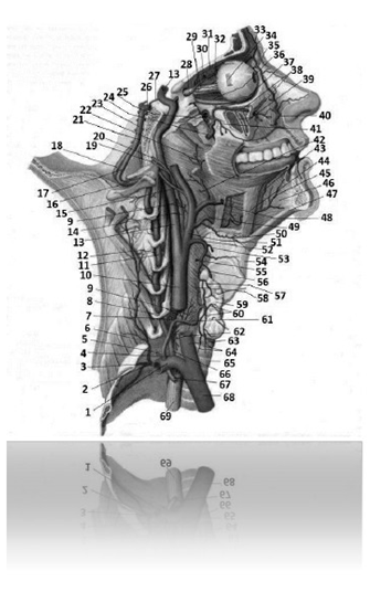
57 – posterior branch of the superior thyroid artery
014 981 564 168
58 – throat 519 987 319 427
59 – thyroid gland 829 319 409 819
60 – glandular branches 149 816 013 009
61 – inferior thyroid artery 518 377 918 478
62 – esophageal branches 519 512 319 812
63 – common carotid artery 894 317 212 847
64 – tracheal branches 919 810 499 310
65 – trachea 429 318 919 888
66 – vertebral artery (prevertebral part)
109 467 219 891
67 – internal thoracic artery 598 341 818 941
68 – brachiocephalic trunk 998 301 248 227
69 – esophagus 598 381 698 711
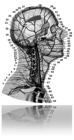
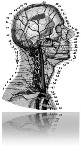
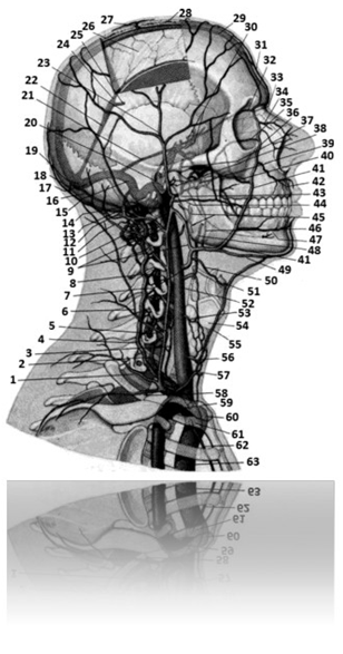
Fig. 91 Veins of the head and neck (right view)
598 716 319 816:
1 – transverse vein of the neck 814 416 214 319
2 – vertebral vein 146 472 019 541
3 – anterior vertebral vein 109 516 918 416
4 – accessory vertebral vein 210 341 907 654
5 – external jugular vein 594 716 814 516
6 – deep vein of the neck 801 498 548 617
7 – facial vein 599 715 819 316
8 – external vertebral venous plexuses
421 054 329 891
9 – submandibular vein 364 817 384 199
10 – superior jugular bulb 448 546 891 479
11 – occipital vein 914 712 298 267
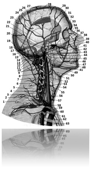
12 – condylar emissary vein 648 513 694 817
13 – posterior auricular vein 368 198 549 617
14 – mammillary emissary vein 391 849 501 011
15 – sigmoid sinus 109 516 397 894
16 – occipital sinus 012 126 094 791
17 – transverse sine 549 716 398 471
18 – occipital emissary vein 548 617 294 581
19 – sine drain 364 814 501 122
20 – inferior petrosal sinus 284 368 149 017
21 – superior petrosal sinus 019 596 394 717
22 – direct sine 849 712 646 181
23 – superficial temporal veins 694 513 814 216
24 – inferior sagittal sinus 814 316 219 497
25 – great cerebral vein 498 142 549 617
26 – falx cerebri 001 918 021 378
27 – parietal emissary vein 349 161 894 717
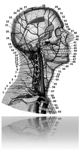
28 – diploic veins 148 564 219 617
29 – superior sagittal sinus 914 715 514 292
30 – cavernous sinus 846 139 948 581
31 – supratrochlear vein 819 621 398 471
32 – superior ophthalmic vein 909 610 549 798
33 – nasofrontal vein 514 369 129 710
34 – external nasal vein 319 481 589 671
35 – inferior ophthalmic vein 149 678 148 591
36 – angular vein 693 146 590 310
37 – middle meningeal vein 018 531 219 641
38 – veins of the parotid gland 516 949 140 510
39 – pterygoid plexus 591 248 791 260
40 – deep facial vein 309 864 194 971
41 – facial vein 599 715 819 316
42 – superior labial vein 504 361 309 584
43 – maxillary vein 598 314 818 914
44 – transverse facial vein 698 713 294 167
45 – Pharyngeal veins 019 818 594 614
46 – palatine vein 548 316 819 471
47 – inferior labial vein 547 218 599 641
48 – lingual vein 318 586 389 471
49 – submental vein 194 368 594 817
50 – superior thyroid vein 648 471 201 199
51 – hyoid bone 549 316 219 841
52 – internal jugular vein 598 612 719 322
53 – middle thyroid vein 814 017 201 849
54 – thyroid cartilage 588 421 388 711
55 – anterior jugular vein 368 541 291 479
56 – inferior thyroid vein 108 641 294 719
57 – inferior bulb of the jugular vein 512 621 221 848
58 – suprascapular vein 548 571 818 548
59 – subclavian vein 598 317 898 214
60 – brachiocephalic vein (left) 219 378 919 278
61 – brachiocephalic vein (right) 219 378 919 278
62 – internal thoracic vein 491 316 894 198
63 – superior vena cava 398 712 988 012
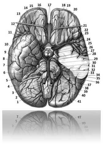
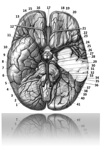
Fig. 92 Arteries of the brain (bottom view)
498 641 898 478:
1 – branch of the cerebellar tonsil 549 641 891 748
2 – posterior inferior cerebellar artery 469 718 549 641
3 – hypoglossal nerve 548 321 555 678
4 – cranial nerve 584 647 289 741
5 – anterior inferior cerebellar artery 598 691 798 641
6 – piece 897 567 971 319
7 – choroid plexus of the fourth ventricle
694 167 298 547
8 – arteries of the bridge 467 589 196 318
9 – superior cerebellar artery 361 948 594 161
10 – oculomotor nerve 519217519217
11 – optic tract 519 218 919 245
12 – hypothalamic infundibulum 519 211 919 000
13 – optic chiasm 010 216 319517
14 – olfactory triangle 518 642 319 716
15 – olfactory tract 718 217 458 917
16 – Olfactory bulb 024 312 598 742
17 – anterior cerebral arteries (postcommunication)
part) 468 514 398 617
18 – medial orbitofrontal branch 518 641 201 009
19 – anterior communicating artery 641 478 594 641
20 – anterior cerebral arteries (precommunication)
part) 584 061 412 011
21 – lateral frontobasal artery 618 531 214 712
22 – internal carotid artery 549 712 810 248
23 – insular arteries 518 714 316 214
24 – middle cerebral artery 496 491 817 514
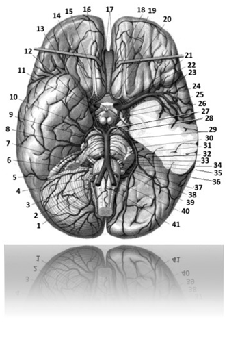
25 – anterior villous artery of the choroid plexus
491 316 498 714
26 – posterior communicating artery 548 641 298 781
27 – posterior cerebral artery (precommunicating)
part) 548 641 798 521
28 – posterior cerebral artery (postcommunicating)
part) 316 594 218 749
29 – basilar artery 851 478 594 814
30 – trigeminal nerve 549 319 818 711
31 – abducens nerve 514 517 214 812
32 – posterior cerebral artery (temporal part)
498 671 291 491
33 – intermediate nerve 219 381 648 719
34 – facial nerve 999 811 319 211
35 – vestibulocochlear nerve 548 217 918 421
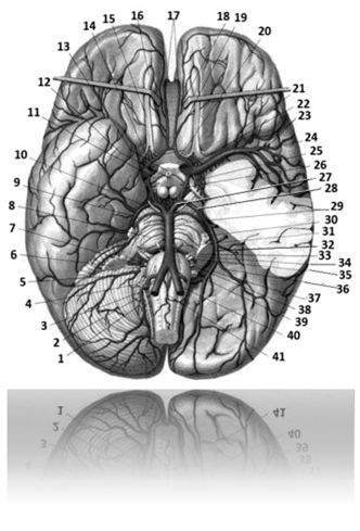
36 – lateral occipital artery (terminal part)
619 012 504 794
37 – medial occipital artery (terminal part)
581 704 916 219
38 – glossopharyngeal nerve 519 371 214 572
39 – vagus nerve 489 981 728 221
40 – vertebral artery (left) (intracranial part)
364 819 498 471
41 – anterior spinal artery 617 281 707 914
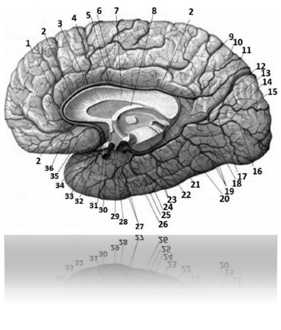
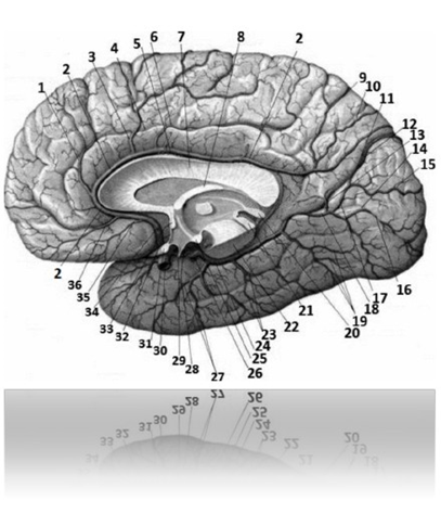
Fig. 93 Arteries of the brain (medial
surface) 469 518 716 491:
1 – intermediate-medial frontal branch
498 617 319 478
2 – anterior cerebral artery 317 498 689 171
3 - posteromedial frontal branch (anterior
cerebral artery) 781 496 319 641
4 – cingulate groove 579 312 919 021
5 – groove of the corpus callosum 248 312 848 212
6 – cingulate branch (anterior cerebral artery)
497 101 398 712
7 – corpus callosum 498 712 328 071
8 – vault 648 314 589 716
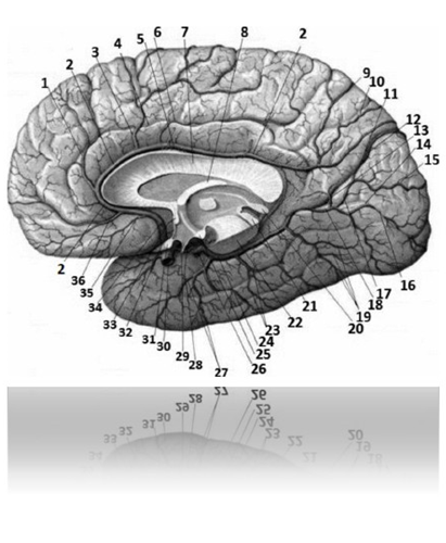
9 – paracentral branch (anterior cerebral
arteries) 364 817 294 317
10 – preclinical branch (anterior cerebral artery)
694 171 894 214
11 – parieto-occipital groove 691 378 549 617
12 – parieto-occipital branch (posterior cerebral
arteries) 691 314 291 491
13 – parietal branch (posterior cerebral artery)
814 216 497 218
14 – occipitotemporal branch (posterior cerebral
arteries) 514 618 319 481
15 – calcarine branch (posterior cerebral artery)
564 814 219 417
16 – spur groove 214 318 414 888
17 – posterior cerebral artery 594 361 809 491
18 – medial occipital artery 581 704 916
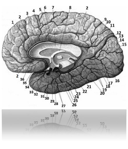
19 – posterior temporal branches (posterior cerebral
arteries) 649 371 298 481
20 – pineal gland (epiphysis) 489 641 399 042
21 – roof plate (quadrigeminal plate)
514 317 818 212
22 – brain stem 918 412 818 212
23 – intermediate temporal branches 549 648 391 361
24 – lateral occipital artery 496 819 716 478
25 – mammillary body 534 817 214 712
26 – posterior cerebral artery (temporal part)
498 671 291 491
27 – anterior temporal branches (lateral
occipital artery) 648 531 219 471
28 – posterior communicating artery 548 641 298 781
29 – deepening of the funnel at the bottom of the third brainstem
ventricle 568 016 219 014
30 – internal carotid artery 549 712 810 248
31 – optic chiasm 010 216 319 517
32 – terminal plate 048 541 298 647
33 – anterior commissure 389 691 974 846
34 – anterior communicating artery
641 478 594 641
35 – medial frontobasal artery
(medial orbitofrontal branch)
518 641 201 009
36 – anteromedial frontal branch (anterior
cerebral artery) 698 142 489 716
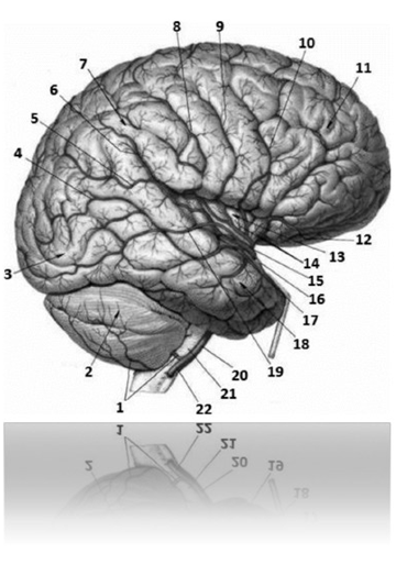
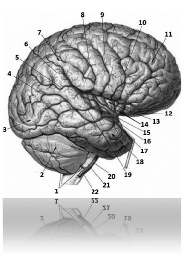
Fig. 94 Arteries of the brain
(superolateral surface)
469 471 898 417:
1 - posterior inferior cerebellar artery
469 718 549 641
2 – cerebellum 828 219 328 299
3 – occipital lobe 519 617 298 714
4 – artery of the angular gyrus 519 617 289 741
5 – posterior parietal artery 164 816 319 471
6 – anterior parietal artery 014 146 214 814
7 – parietal lobe 618 041 894 141
8 – artery of the postcentral sulcus
102 348 410 514
9 – artery of the central sulcus 648 318 489 316
10 – artery of the precentral groove
584 216 218 714
11 – frontal lobe 316 618 319 417
12 – lateral frontobasal artery
618 531 214 712
13 – islet 584 216 298 741
14 – insular arteries 518 714 316 214
15 – middle cerebral artery 496 491 817 514
16 – anterior temporal artery 689 371 298 681
17 – middle temporal artery 618 591 294 317
18 – temporal lobe 564 931 298 714
19 – posterior temporal artery 548 721 296 397
20 – basilar artery 851 478 594 814
21 – bridge 248 317 284 271
22 – vertebral artery (right) (intracranial
part) 364 819 498 471
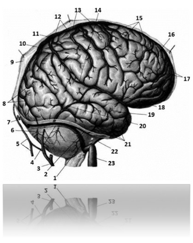
Fig. 95 Superficial cerebral veins
(superolateral surface)
649 712 519 747:
1 – sigmoid sinus 109 516 397 894
2 – superior petrosal sinus 019 596 394 717
3 – posterior auricular vein 368 198 549 617
4 – transverse sine 549 716 398 471
5 – occipital vein 914 712 298 267
6 – mammillary emissary vein 391 849 501 011
7 – occipital emissary vein 548 617 294 581
8 – occipital veins 789 621 298 491
9 – dura mater of the brain
333 489 312 289
10 – parietal lobe 618 041 894 141
11 – lateral venous lacuna 569 317 398 471
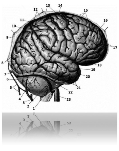
12 – parietal vein 394 569 398 741
13 – superior sagittal sinus 914 715 514 292
14 – superior anastomotic vein 341 318 519 641
15 – frontal veins 361 384 219 471
16 – frontal lobe 316 618 319 417
17 – prefrontal veins 598 641 298 791
18 – superficial middle cerebral vein
694 178 394 516
19 – temporal lobe 564 931 298 714
20 – inferior anastomotic vein 391 568 914 918
21 – inferior cerebral veins 719 628 514 318
22 – inferior petrosal sinus 284 368 149 017
23 – internal jugular vein 598 612 719 322
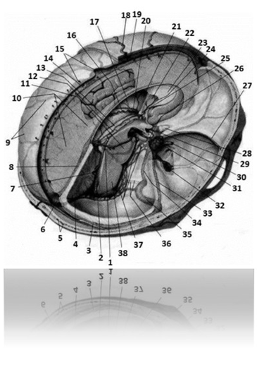
Fig. 96 Sinuses of the dura mater
691 958 549 164:
1 – basal vein 202 464 891 319
2 – great cerebral vein 498 142 549 617
3 – transverse sine 549 716 398 471
4 – occipital sinus 012 126 094 791
5 – diploic veins 148 564 219 617
6 – sine drain 364 814 501 122
7 – fourth ventricle 514 321 414 218
8 – direct sine 849 712 646 181
9 – superior cerebral veins 519 316 489 718
10 - large sickle-shaped process of the dura
meninges 364 815 398 574
11 – bridge 248 317 284 271
12 – internal cerebral vein 541 219 319 471
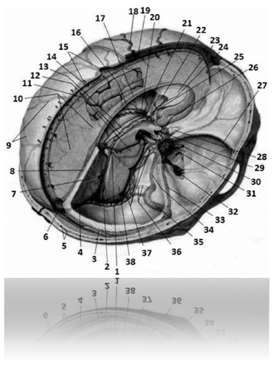
13 – vascular basis of the third ventricle 584 217 284 917
14 – superior sagittal sinus 914 715 514 292
15 – lateral lacunae 398 741 298 474
16 – superior villous vein 518 361 987 241
17 – inferior sagittal sinus 814 316 219 497
18 – superficial temporal vein 548 327 918 227
19 – parietal emissary vein 349 161 894 717
20 – superior thalamostriate veins 598 164 398 711
21 – lateral ventricle 649 140 508 914
22 – transparent septum plate
319 798 549 164
23 – genu of the corpus callosum 148 512 319 417
24 – anterior vein of the septum pellucidum
849 617 219 514
25 – vault pillar 168 794 598 716
26 – intercavernous sinuses 514 019 598 411
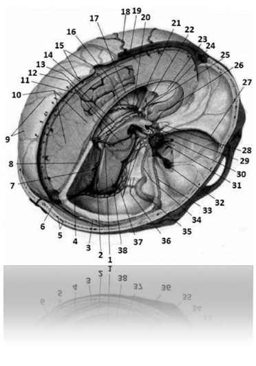
27 – sphenoparietal sinus 514 312 619 718
28 – optic nerve 448 817 918 217
29 – internal carotid artery 549 712 810 248
30 – superficial middle cerebral vein
694 178 394 516
31 – cavernous sinus 846 139 948 581
32 – venous plexus of the oval opening
498 716 374 917
33 – superior petrosal sinus 019 596 394 717
34 – inferior petrosal sinus 284 368 149 017
35 – sigmoid sinus 109 516 397 894
36 – venous plexus of the hypoglossal canal
598 611 998 164
37 – basilar plexus 691 319 819 471
38 – medulla oblongata 514 417 814 217
Arteries and veins of the heart 514 814 219 417
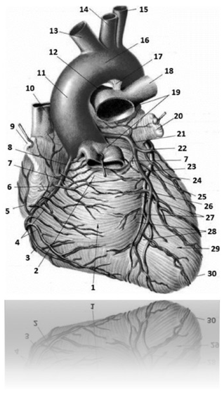
Fig. 97 Arteries and veins of the heart (sternocostal
surface) 514 814 219 417:
1 – right ventricle of the heart 598 371 988 011
2 – arterial cone 548 647 219 741
3 – right marginal branch of the right coronary artery
518 617 219 419
4 – anterior cardiac veins 648 715 594 317
5 – intermediate atrial branches
594 781 978 471
6 – coronal groove 519 312 814 829
7 – branch of the arterial cone 394 168 574 971
8 – right coronary artery 691 368 519 479
9 – right cardiac auricle 598 714 321 898
10 – superior vena cava 398 712 988 012
11 – ascending aorta 598 712 898 612
12 – right pulmonary artery 694 897 594 716
13 – brachiocephalic trunk 998 301 248 227
14 – common carotid artery (left) 428 712 488 913
15 – subclavian artery (left) 429 387 219 377
16 – aortic arch 219 877 549 277
17 – arterial ligament 519 481 319 818
18 – left pulmonary artery 691 318 497 541
19 – pulmonary trunk 519 421 819 221
20 – left cardiac auricle 519 318 219 481
21 – left coronary artery 194 641 291 891
22 – circumflex branch of the left coronary artery
619 471 218 514
23 – left anterior interventricular branch
coronary artery 364 891 291 471
24 – left marginal branch 589 714 210 481
25 – great cardiac vein 641 217 498 718
26 – lateral branch 491 316 218 714
27 – septal interventricular branches
691 314 219 718
28 – anterior interventricular groove
909 817 398 787
29 – left ventricle 589 348 914 918
30 – apex of the heart 519 421 899 321
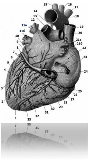
Fig. 98 Arteries and veins of the heart (diaphragmatic
surface) 514 816 914 317:
1 – apex of the heart 519 421 899 321
2 – left ventricle 589 348 914 918
3 – anastomotic atrial branch
614 917 918 517
4 – left marginal branch 589 714 210 481
5 – posterior vein of the left ventricle 429 318 719 888
6 – posterior branch of the left ventricle 467 548 219 741
7 – circumflex branch of the left coronary artery
619 471 218 514
8 – great cardiac vein 641 217 498 718
9 – oblique vein of the left atrium 598 714 319 814
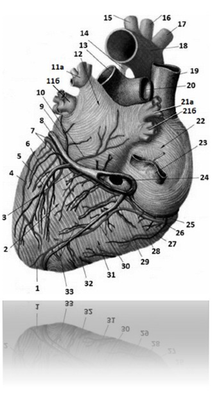
10 – intermediate atrial branches
594 781 978 471
11a – left superior pulmonary vein 549 671 298 471
11b – left inferior pulmonary vein 598 649 219 714
12 – left atrium 518 712 314 887
13 – left pulmonary artery 697 108 889 491
14 – arterial ligament 519 481 319 818
15 – subclavian artery (left) 429 387 219 377
16 – common carotid artery (left) 428 712 488 913
17 – brachiocephalic trunk 998 301 248 227
18 – aortic arch 219 877 549 277
19 – superior vena cava 398 712 988 012
20 – right pulmonary artery 694 897 594 716
21a – right superior pulmonary vein 691 894 319 712
21b – right inferior pulmonary vein 697 213 519 491
22 – right atrium 491 016 519 497
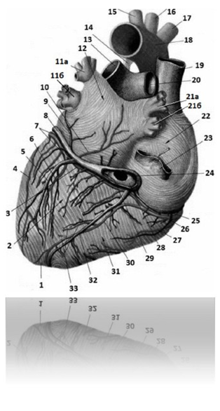
23 – inferior vena cava 549 671 919 871
24 – opening of the coronary sinus 548 641 219 712
25 – small cardiac vein 598 712 918 322
26 – right coronary artery 691 368 519 479
27 – right posterolateral branch 479 691 319 814
28 – coronary sinus valve 584 316 219 479
29 – coronary sinus of the heart 578 916 219 316
30 – posterior interventricular branch of the right coronary artery
arteries 589 718 549 641
31 – right ventricle of the heart 598 371 988 011
32 – middle cardiac vein 589 641 298 791
33 – septal interventricular branches
691 314 219 718
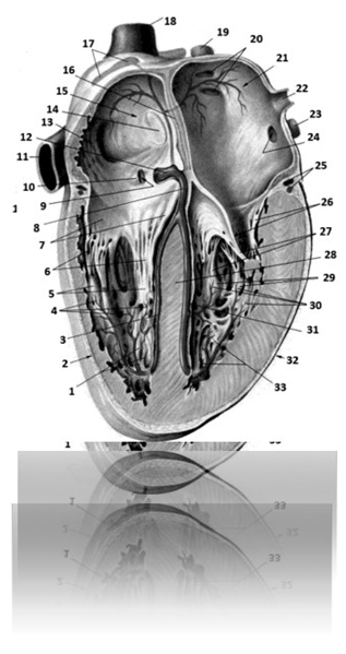
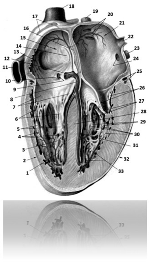
Fig. 99 Conduction system of the heart
989 808 884 318:
1 – fleshy trabeculae 194 217 289 678
2 – right ventricle of the heart 598 371 988 011
3 – right branch of the atrioventricular bundle
319 728 549 641
4 – subendocardial branches of the right ventricle
641 218 514 017
5 – papillary muscles of the right ventricle
491 318 597 317
6 – tendinous chordae of the tricuspid valve
641 318 219 748
7 – right atrioventricular (tricuspid)
(chat) valve 389 412 819 322
8 – atrioventricular bundle of His
198 316 398 714
9 – opening of the coronary sinus 548 641 219 712
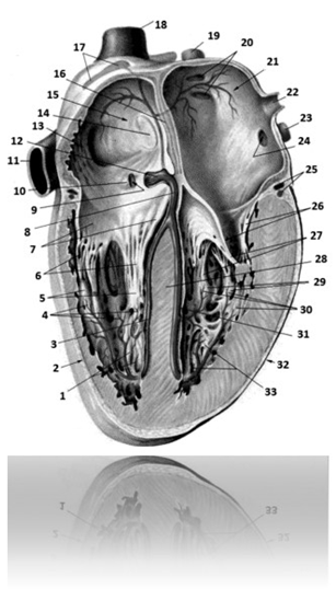
10 – coronary sinus valve of the heart
584 316 219 479
11 – inferior vena cava 549 671 919 871
12 – atrioventricular node 169 381 219 714
13 – pectineus muscles 391 689 598 714
14 – oval fossa 394 169 519 718
15 – right atrium 491 016 519 497
16 – interatrial septum 894 158 019 617
17 – sinus node 368 491 298 749
18 – superior vena cava 398 712 988 012
19 – right superior pulmonary vein 691 894 319 712
20 – openings of the pulmonary veins 589 671 298 491
21 – left atrium 518 712 314 887
22 – left superior pulmonary vein (part of the figure)
549 671 298 471
23 – left inferior pulmonary vein 598 649 219 714
24 – opening of the lower left pulmonary vein
498 712 219 714
25 – vessels of the heart 549 648 519 716
26 – left atrioventricular (mitral)
valve 598 517 818 617
27 – mitral valve tendinous chordae
794 814 519 714
28 – interventricular septum 548 581 218 491
29 – papillary muscles of the left ventricle
467 219 519 712
30 – subendocardial branches of the left ventricle
497 518 584 718
31 – left branch of the atrioventricular bundle
(Hiss bundle) 649 191 218 549
32 – left ventricle of the heart 589 348 914 918
33 – fleshy trabeculae of the left ventricle
468 791 298 745
Veins of the lower extremities 589 712 319 614
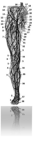
Fig. 100 Veins of the lower extremities 589 712 319 614:
1 – dorsal venous network of the foot 197 298 108 641
2 – dorsal venous arch of the foot 194 389 794 216
3 – venous network of the leg 784 594 316 497
4 – anterior tibial veins 589 714 319 718
5 – knee veins 817 316 368 491
6 – small saphenous vein of the leg 169 381 379 149
7 – venous network of the thigh 497 581 369 794
8 – deep vein of the thigh 184 517 396 847
9 - lateral veins surrounding the femur
649 132 389 714
10 – superficial vein encircling the iliac bone
317 849 178 471
11 – superficial epigastric vein 491 694 218 713
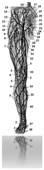
12 – external iliac vein 999 888 719 898
13 – deep circumflex iliac vein
721 394 549 718
14 – iliolumbar veins 548 791 018 216
15 – lumbar vein 589 712 919 261
16 – inferior vena cava 549 671 919 871
17 – common iliac vein 548 713 918 781
18 – median sacral vein 598 717 318 917
19 – lateral sacral veins 549 316 298 711
20 – internal iliac vein 549 316 814 787
21 – sacral venous plexus 649 271 298 541
22 – inferior gluteal veins 497 189 369 141
23 – internal pudendal vein 641 894 594 818
24 – obturator vein 318 414 849 161
25 – external genital vein 648 143 019 549
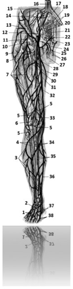
26 – superficial dorsal veins of the penis
314 647 217 498
27 – anterior scrotal veins 541 314 819 317
28 – medial veins surrounding the femur
364 981 219 784
29 – additional saphenous vein of the leg 369 489 598 716
30 – perforating vein 414 548 374 811
31 – great saphenous vein of the leg 496 794 894 175
32 – femoral vein 316 584 912 848
33 – popliteal vein 149 721 801 497
34 – peroneal veins 518 364 194 816
35 – posterior tibial veins 479 514 194 817
36 – great saphenous vein of the leg 496 794 894 175
37 – dorsal metatarsal veins of the foot 648 564 817 219
38 – dorsal digital veins of the foot 197 248 594 714
Arteries, veins and capillaries 219 387 919 887
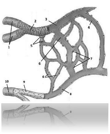
Fig. 101 Microcirculatory bed 549 318 497 561:
1 – artery 894 547 284 717
2 – arteriole 694 574 895 601
3 – precapillary 608 491 298 491
4 – arteriolo-venular anastomosis 549 316 897 314
5 – precapillary sphincters 649 172 218 371
6 – capillaries 479 821 294 364
7 – capillary sphincters 185 494 016 001
8 – postcapillary101 498 754 361
9 – venule 669 517 918 394
10 – Vienna 149 621 818 318
CENTRAL NERVOUS SYSTEM (continued)
291 384 074 217
Brain 814 729 318 818
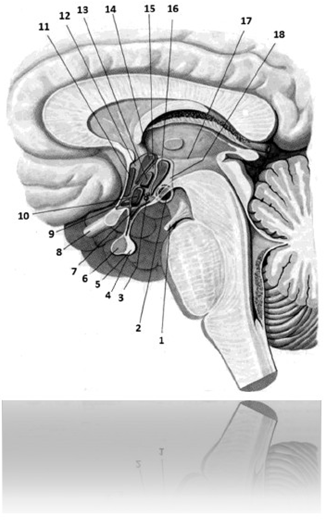
Fig. 102 Hypothalamus 918 671 818 971
Pituitary gland 317 218 219 819:
1 – oculomotor nerve 519 217 519 217
2 - medial and lateral nuclei of the mammillary
bodies 498 716 219 418
3 – gray-humped cores 898 141 218 718
4 – pituitary gland 317 218 219 819
5 – neurohypophysis 814 061 217 041
6 – adenohypophysis (anterior lobe) 518 041 219 498
7 – funnel core (arcuate core) 497 581 264 714
8 – optic nerve 448 817 918 217
9 – supraoptic nucleus 948 516 714 271
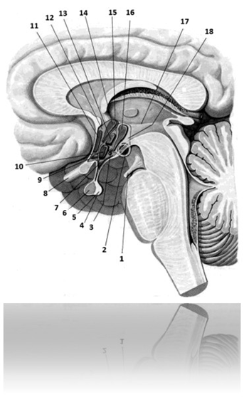
10 – inferior medial hypothalamic nucleus
178 491 219 617
11 – preoptic nucleus 718 471 219 648
12 – anterior hypothalamic region
749 164 218 541
13 – dorsomedial hypothalamic nucleus
549 716 218 541
14 – paraventricular nuclei 598 612 317 491
15 – intermediate hypothalamic region
471 218 549 016
16 – posterior hypothalamic nucleus 001 495 018 547
17 – hypothalamic groove 016 489 808 489
18 – posterior hypothalamic region
018 418 488 518
Content
HEMOPOIESIS AND IMMUNE DEFENSE SYSTEM
219 648 317 918
Central organs of hematopoiesis and immune defense
416 489 319 641
Peripheral organs of hematopoiesis and immune defense
794 916 219 481
Unified immune system of the mucous membranes
674 981 219 496
BLOOD CELLS 549681219717
Leukocytes 694 218 574 271
Agranulocytes 548 274 298 641
Granulocytes 918 547 219 714
DENTOALVEOLAR SYSTEM 216 548 219 716
Bones of the facial part of the skull 219 715 819 815
Teeth 698 314 819 516
Organs of the oral cavity
Chewing and facial muscles
Temporomandibular joint
Glands of the vestibule and oral cavity 498 617 219 491
SPINE. JOINTS, LIGAMENTS AND MUSCLES OF THE SPINE
Vertebral column 214 217 000 819
VERTEBRAE 498 641 319 048
Vertebral motor segment 714 986 219 694
Muscles and ligaments of the spine 549 641 894 217
LIGAMENTS OF THE PELVIS AND HIP JOINT 498 641 798 478
MUSCLES AND FASCIA OF THE BACK AND OCCIUM 798 041 261 509
FEMALE GENITAL ORGANS 519 814 089 319
Female external genitalia 519 319 818 678
Internal female genital organs 419 219 808 319
MAMMARY GLAND 648317219491
CARDIOVASCULAR SYSTEM
Arteries and veins of the heart 514 814 219 417
Veins of the lower extremities 589 712 319 614
Arteries, veins and capillaries 219 387 919 887
CENTRAL NERVOUS SYSTEM (continued)
291 384 074 217
Brain 814 729 318 818