
INTRODUCTION
When restoring human matter with numerical concentrations, the following methods can be used:
1) Read the numbers of the series corresponding to the restored matter, written after the name of the matter;
2) Mentally pronounce the numbers of the series corresponding to the restored matter;
3) Look at the image or name of the matter and mentally pronounce the numbers of the corresponding row;
4) Imagine that you are between the numbers of the series corresponding to the matter being recreated, having a large size. The numbers between which you imagine yourself, you need to strive to perceive distinctly. The light from these numbers can reach you. The above actions can be performed with any numbers in the series.
5) Imagine that you are looking at a number series from above.
6) Represent a numerical series in the area that you are restoring. To do this, you need to use the image of matter given in this book, relating to the number series you are using.
7) Represent the numerical series between the image of matter and part of the mirror image given in this book that relate to the number series used.
8) By comparing the numbers of the series, you can determine the relationship between the various human matter that controls the norm. It is possible to restore matter using a series of other matter. In this case, when using the numerical series of the matter that is being restored, you can simultaneously or sequentially use the numerical series of other matter. From the numerical series of other matter, you can first concentrate on the numbers that coincide with the numbers of the series of the matter being restored. Then you can use the entire numerical series of other matter, mentally drawing a light beam from it that intersects the numerical series of the matter or the matter itself that you are restoring.
When perceiving a rapid restorative effect, you can determine the next point or area in the organism after the matter being restored itself, through which the restored matter is created. This next point or area will then be in other matter, through whose numerical series the restoration of the matter you have chosen is carried out. There may be many subsequent points or areas through which the creation of matter is carried out. The first point or area of creation of the selected matter is in the matter itself.
Having established the points or areas of creation of the restored matter by using numerical series, it is possible to restore the matter by concentrating on these points or areas. In this case, a spiritual state is established that corresponds to the restoration and norm of the selected matter.
Remembering and establishing the indicated state of the spirit, it is possible to restore the matter with the spirit, which is thus life-giving. Then it is possible to extend such spiritual action to all the matter of the organism, taking into account external events, and thereby achieve a spiritual state that corresponds to eternal development. In certain cases, depending on the angle of perception, different numerical series may correspond to the restored matter.
9) To speed up the restoration of human matter, gaps in numerical series can be perceived as gaps between words in a sentence. Then, for each numerical component of the entire series, separated by a space, you can consider the word that means the normal functioning of matter, to which the series corresponds. Creator the level that creates matter corresponding to the numerical series, and the matter of the whole organism can be perceived, trying to perceive such a word. Light, creating matter corresponding to the numerical series, spreads according to the laws of optics on all other matter of the human body and on the environment. Here you can understand why Some sensations and emotions are perceived as external.
This allows for more accurate recognition of where on the level
event management must act on the basis of the interaction of body tissues and where on the basis of interactions of the body's matter and the environment. This method of precise recognition allows more effectively manage events to the level of the normal state of the body's matter, regardless of any circumstances.
With this method, you simultaneously perceive the tissue of the body and the events surrounding the person. as if you were looking at what was described with your physical sight. And depending on the situation, you can take solution – how to act in the direction of eternal development. In some cases, you can take physical actions and in some periods to produce spiritual action to normalize events in the direction of eternal life.
This perception of yours develops your spirit, soul and physical body to the level at which creation of human matter occurs on a spiritual basis. Numbers allow us to obtain an accurate spiritual state, corresponding to the norm of human matter. To strengthen control, you can use the well-known, that is, knowledge from physics about the corpuscular-wave duality of matter, widely entrenched in the collective consciousness, according to which any object can exhibit both wave properties and the properties of a particle of matter.
Creating by concentrating on the number series of light waves corresponding to the norm of human matter, you create
normally functioning human matter.
All data in this book on the methods of restoring human matter using numerical concentrations can be use for preventive health purposes, for rejuvenation, if necessary to restore matter, and regardless of the initial data from which the matter is restored.
When using the specified In points 1 through 9 of the introduction of methods, the following can be taken into account:
1. For preventive purposes, it is advisable to carry out health improvement with the simultaneous distribution the effects of numerical concentrations on the future.
2. When rejuvenating, it is advisable to first consistently concentrate on the number series, located in the content, taking into account the task of eternal development, then concentrate on the matter that you
locally rejuvenate.
3. By restoring the body's matter, it is possible to produce numerical concentrations of successively different given in the book in the ways. In this case, it is possible to use both the numerical series corresponding to the reconstructed matter, as well as the numerical series of the area that includes the matter that you are restoring.
4. If it is necessary to restore matter after biological death, then one should concentrate on numbers first sequentially from left to right, then in reverse order from right to left.
The spiritual impulse that creates human matter allows for the expansion of recovery methods.
In restoring the matter of man, one must strive to develop the spiritual level to a state in which human matter is created and functions by spiritual action along with biological and event-based
principles.
Such a spiritual state, when implementing the methods of eternal development, should ensure complete restoration of human matter regardless of initial data and any circumstances.
CELLS AND TISSUES 829 379 429 841
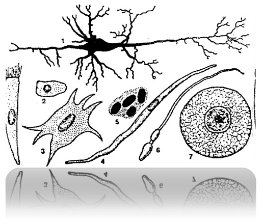
Fig. 1 Cell shapes
1 – nervous 519 513 819 814
2 – epithelial 518 321 678 024
3 – connective tissue 819 417 319 814
4 – smooth muscle 519 312 419 814
5 – erythrocyte 214 719 319 818
6 – sperm 319 814 888 918
7 – egg 888 319 914 718
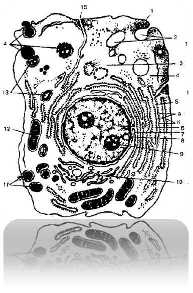
Fig. 2 Ultramicroscopic structure of a cell
1 – cytolemma (plasma membrane) 814 718 314 213
2 – pinocytotic vesicles 214 718 314 218
3 – centrosome (cell center, cytocenter) 519 217 018 017
4 – hyaloplasm 614 217 321 218
5 – endoplasmic reticulum 819 517 319 418 (a – membranes
endoplasmic reticulum, b – ribosomes)
6 – core 814 321 718 912
7 – connection of the perinuclear space with cavities
endoplasmic reticulum 819 421 719 378
8 – nuclear pores 918 472 519 318
9 – nucleolus 918 412 718 814
10 – intracellular mesh apparatus (Golgi complex)
819 918 319 217
11 – secretory vacuoles 979 974 348 522
12 – mitochondria 819 317 419 814
13 – lysosomes 519 712 314 518
14 – three successive stages of phagocytosis
819 412 714 321
15 – connection of the cell membrane with membranes
endoplasmic reticulum 514 816 314 819
Fabrics 898 314 988 889
Epithelial tissue 891 389 426 319
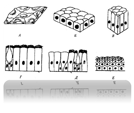
Fig. 3 Different types of epithelium
A – single-layer flat 819 417 319 817
B – single-layer cubic 518 611 918 716
B – cylindrical 318 216 718 916
G - single-layer ciliated 319 821 319 719
D – multi-row 918 216 917 418
E - multilayer keratinizing 428 614 048 910
Connective tissue 719 317 918 517
Muscle tissue 514 312 814 312
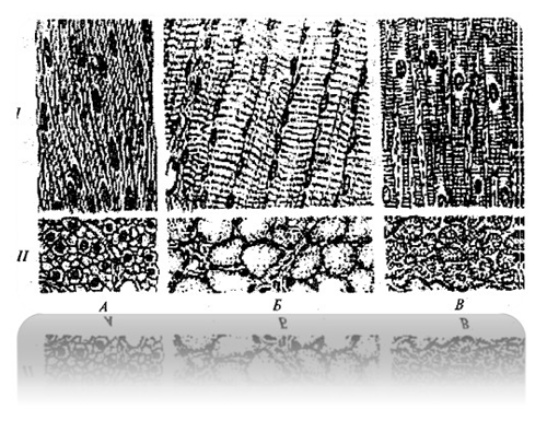
Fig. 4 Types of muscle tissue
I – longitudinal view
II – transverse view
A – smooth (non-striated) 514 718 314 218
B - striated skeletal 917 312 218 412
B – striated cardiac 914 318 514 712
Nervous tissue 718 412 518 914
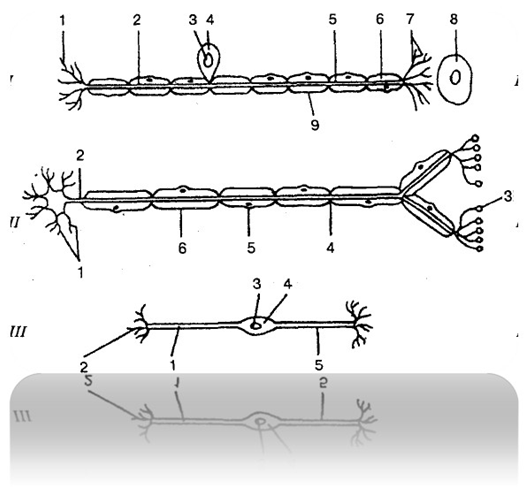
Fig. 5 Structure of a neuron:
I – sensory neuron 814 317 914 918:
1 – neuron endings 519 312 214 712
2 – axon 314 812 219 418
3 – core 314 812 219 217
4 – cell body 917 219 817 519
5 – dendrite 318 517 918 241
6 – myelin sheath 514 717 814 317
7 – receptor 518 214 019 481
8 – organ 814 317 914 817
9 – neurilemma 714 312 814 512
II – motor neuron 319 816 819 312:
1 – dendrites 318 517 918 241
2 – axon 314 812 219 418
3 – end plate 214 217 814 312
4 - Ranvier interception 518 217 818 217
5 – Schwann cell nucleus 214 312 814 212
6 – Schwann cell 841 218 412 518
III – interneuron 314 517 214 817:
1 – axon 314 812 219 418
2 – dendrites 318 517 918 241
3 – core 314 812 219 217
4 – cell body 917 219 817 519
5 – dendron 518 418 719 281
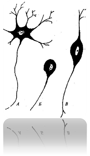
Fig. 6 Types of neurons
A – multipolar 478 641 318 217
B – unipolar 514 371 848 961
B – bipolar 818 217 318 514
NERVOUS SYSTEM 219 317 819 298
CENTRAL NERVOUS SYSTEM 291 384 074 217
Spinal cord 314 218 814 719
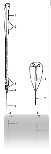
Fig. 7 Spinal cord (diagram):
A:
1 – spinal cord 314 218 814 719
2 – cervical thickening 298 387 984 721
3 – lumbosacral thickening 429 317 219 817
4 – medullary cone 219 381 419 971
5 – end thread 519 317 919 218
B:
1 – terminal ventricle 419 814 218 914
2 – end thread 519 317 919 218
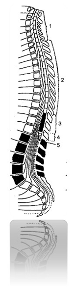
Fig. 8 Spinal cord segments 219 317 888 847
1 – cervical segments (1 – 8) 218 828 204 217,
cervical part 849 218 314 918
2 – thoracic segments (1 – 12), thoracic part 234 891 019 217
3 – lumbar segments (1 – 5), lumbar part 219 004 489 668
4 – sacral segments (1 – 5), sacral part 229 317 916 021
5 – coccygeal segments (1 – 3), coccygeal part 834 219 918 715
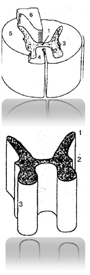
Fig. 9 Spinal cord (diagram)
1 – central channel 888 991 213 451
2 – gray matter 891 021 328 423
3 – white matter 478 217 219 328
4 – front cord 489 219 219 321
5 – lateral cord 290 029 432 517
6 – rear cord 048 217 428 471
Fig. 10. Columns of gray matter of the spinal cord
1 – rear 219 482 319 213
2 – side 214 712 218 521
3 – front 317 484 217 244
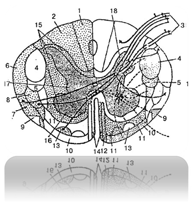

Fig. 11 White matter pathways on the transverse
section of the spinal cord (diagram):
1 – thin beam 219 317 418 217
2 – wedge-shaped bundle 319 215 219 317
3 – posterior root 214 317 219 224
4 – lateral corticospinal (pyramidal) tract
248 317 218 321
5 – rubrospinal tract 428 521 328 721
6 – posterior spinocerebellar tract 888 917 218 912
7 – anterior spinocerebellar tract 918 214 218 217
8 – lateral spinothalamic tract 318 217 218 914
9 – olivospinal tract 514 219 314 819
10 – vestibulospinal tract 219 444 558 913
11 – reticular-spinal tract 219 317 418 213
12 – anterior corticospinal (pyramidal) tract
219 318 214 217
13 – anterior spinothalamic tract 219 317 218 214
14 – tegmental-spinal tract 218 317 219 217
15 – posterior lateral and anterior proper bundles
219 314 214 315
16 – front horn 218 217 314 218
17 – side horn 218 317 214 218
18 – rear horn 489 641 728 591
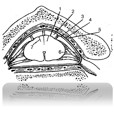
Fig. 12 Spinal cord membranes 219 317 819 312:
1 – pia mater of the spinal cord 849 312 219 814
2 – subarachnoid space 314 712 814 212
3 – arachnoid membrane of the spinal cord 219 317 814 218
4 – dura mater of the spinal cord 218 217 319 218
5 – epidural space 217 314 218 217
6 – serrated ligament 217 318 219 312
7 – intermediate cervical septum 214 321 814 712
Brain 814 729 318 818
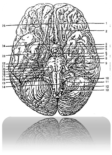
Fig. 13 Base of the brain 219 888 676 021:
1 – olfactory bulb 024 312 598 742
2 – olfactory tract 718 217 458 917
3 – anterior perforated substance 218 317 219 218
4 – gray tubercle 519 317 219 417
5 – optic tract 519 218 919 245
6 – mammillary bodies 534 817 214 712
7 – trigeminal ganglion 418 217 218 217
8 – posterior perforated substance 510 716 519 064
9 – bridge 248 317 284 271
10 – cerebellum 828 219 328 299
11 – pyramid of the medulla oblongata 928 321 728 521
12 – olive 489 217 319 271
13 – spinal nerves 489 218 918 217
14 – hypoglossal nerve 548 321 555 678
15 – accessory nerve 489 917 319 712
16 – vagus nerve 489 981 728 221
17 – glossopharyngeal nerve 519 371 214 572
18 – vestibulocochlear nerve 548 217 918 421
19 – facial nerve 999 811 319 211
20 – abducens nerve 514 517 214 812
21 – trigeminal nerve 549 319 818 711
22 – trochlear nerve 498 064 318 712
23 – oculomotor nerve 519 217 519 217
24 – optic nerve 448 817 918 217
25 – olfactory nerves 478 215 589 315
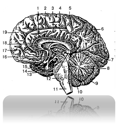
Fig. 14 Brain (sagittal view):
1 – sulcus of the corpus callosum 248 312 848 212
2 – cingulum groove 579 312 919 021
3 – cingulate gyrus 898 312 024 712
4 – corpus callosum 498 712 328 071
5 – central groove 489 213 048 217
6 – paracentral lobule 811 017 319 218
7 – calcarine groove 214 318 414 888
8 – roof plate (quadrigeminal plate)
514 317 818 212
9 – cerebellum 828 219 328 299
10 – IV ventricle 514 321 414 218
11 – medulla oblongata 514 417 814 217
12 – bridge 248 317 284 271
13 – pineal gland (epiphysis) 489 641 399 042
14 – brain stem 918 412 818 212
15 – pituitary gland 317 218 219 819
16 – III ventricle 818 217 418 217
17 – interthalamic fusion 819 312 919 222
18 – transparent partition 214 817 914 817
19 – superior frontal gyrus 918 918 919 217
Medulla oblongata 514 417 814 217

Fig. 15 Medulla oblongata (ventral view):
1 – medulla oblongata 514 417 814 217
2 – back olive field 548 617 319 071
3 – anterior median fissure 598 641 398 216
4 – retro-olive groove 319 217 819 317
5, 9 – anterolateral groove 519 310 819 210
6 – anterior external arcuate fibers 918 217 918 719
7 – pyramid intersection 910 918 819 312
8 – lateral cord 314 912 814 712
10 – olive 489 217 319 271
11 – pyramid of the medulla oblongata 928 321 728 521
Hindbrain 219 317 219 817
Pons and cerebellum 310 564 219 142

1 – cerebellum 828 219 328 299
2 – cerebellopontine triangle 214 312 814 912
3 – bulbar-bridge groove 819 312 219 217
4 – basilar groove 519 312 219 212
5 – middle cerebellar peduncle 219 710 819 210
6 – bridge 248 317 284 271
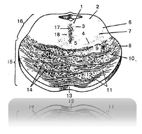
Fig. 17 Cross-section view of the bridge (diagram):
1 – posterior longitudinal fasciculus 519 317 819 213
2 – midbrain tract of the trigeminal nerve 519 017 819 317
3 – medial longitudinal fasciculus 517 918 917 819
4 – medial loop 319 021 713 211
5 – reticular formation 519 312 919 812
6 – trigeminal loop (trigeminothalamic tract)
819 417 919 817
7 – spinal cord loop 514 312 914 212
8 – pontocerebellar fibers 519 471 919 012
9 – corticonuclear fibers 519 315 219 818
10 – cortical-pontine fibers 398 641 988 042
11 – bridge cores 514 312 914 214
12 – corticospinal fibers 218 319 219 418
13 – basilar groove 519 312 219 212
14 – transverse fibers of the bridge 519 312 919 218
15 – anterior (basilar) part of the bridge 588 217 918 214
16 – rear part of the bridge (bridge cover) 819 312 919 212
17 – bridge seam 319 481 498 671
18 – tegmental-spinal tract 518 217 918 217
Midbrain 519 417 819 210
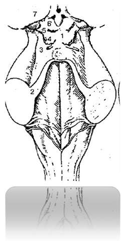
Fig. 18 Midbrain and rhomboid fossa 519 317 888 910:
1 – roof plate (quadrigeminal plate) 514 317 818 212
2 – superior cerebellar peduncle 219 716 219 511
3 – triangle loop 519 217 314 717
4 – lower mound 514 817 914 917
5 – upper mound 918 071 518 971
6 – handle of the lower mound 519 217 918 201
7 – handle of the upper mound 519 322 068 290
Diencephalon 919 213 819 223
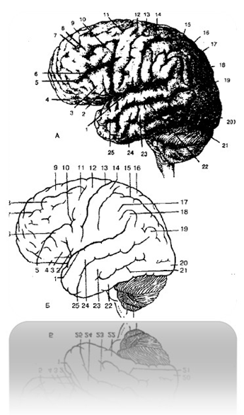
Fig. 19 Brain: superolateral surface,
grooves and convolutions (diagram):
A, B:
1 – lateral groove 514 213 814 213
2 – tegmental part of the inferior frontal gyrus 219 312 819 222
3 – triangular part of the inferior frontal gyrus 888 917 918 217
4 – orbital part of the inferior frontal gyrus 498 641 398 071
5 – inferior frontal groove 498 213 718 223
6 – inferior frontal gyrus 519 312 519 212
7 – superior frontal groove 514 317 514 817
8 – middle frontal gyrus 514 017 314 917
9 – superior frontal gyrus 918 918 919 217
10 – lower precentral groove 519 217 919 817
11 – superior precentral groove 918 512 318 412
12 – precentral gyrus 479 318 012 891
13 – central groove 489 213 048 217
14 – postcentral groove 519 217 918 227
15 – intraparietal groove 418 218 219 371
16 – superior parietal lobule 514 318 914 818
17 – inferior parietal lobule 514 319 214 819
18 – supramarginal gyrus 918 883 518 913
19 – angular gyrus 519 311 918 911
20 – occipital pole 519 317 819 517
21 – inferior temporal sulcus 519 217 219 317
22 – inferior temporal gyrus 519 218 919 519
23 – middle temporal gyrus 519 712 919 212
24 – superior temporal gyrus 518 317 918 217
25 – superior temporal sulcus 514 219 314 919
Ventricles of the brain 989 061 714 318
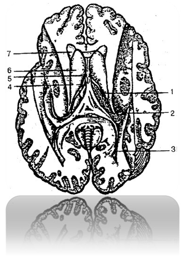
Fig. 20 Lateral ventricles of the cerebrum 919 814 919 217
1 – central part of the lateral ventricle 248 614 319 641
2 – lower horn 319 817 919 917
3 – rear horn 798 617 398 541
4 – interventricular opening 548 321 918 811
5 – transparent partition 214 817 914 817
6 – head of the caudate nucleus 511 064 918 244
7 – front horn 514 312 518 212
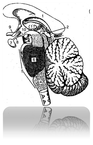

Fig. 21 Rhombencephalon (sagittal view)
314 617 918 554:
1 – corpus callosum 498 712 328 071
2 – pineal body (epiphysis) 489 641 399 042
3 – cerebellum 828 219 328 299
4 – medulla oblongata 514 417 814 217
5 – rhombencephalon 314 617 918 554
6 – bridge 248 317 284 271
7 – pituitary gland 317 218 219 819
8 – funnel 519 211 919 000
9 – optic chiasm 010 216 319 517
10 – hypothalamus 918 671 818 971
11 – interthalamic fusion 248 719 361 989
Meninges of the brain 519 317 819 217
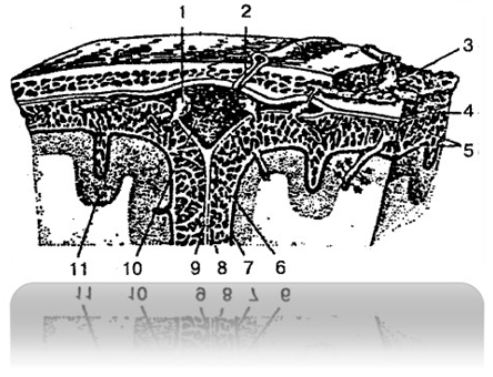
Fig. 22 Meninges of the brain 519 317 819 217:
1 – granulation of the arachnoid membrane 519 617 888 918
2 – emissary vein 519 012 919 722
3 – vein of spongy bone 518 712 318 222
4 – dura mater 333 489 312 289
5 – arachnoid membrane crossbars 519 317 919 227
6 – subarachnoid space 318 271 228 971
7 – choroid 514 322 814 212
8 – arachnoid membrane 319 217 064 827
9 – falx cerebri 001 918 021 378
10 – superior sagittal sinus 914 715 514 292
11 – cerebral cortex 918 617 619 017
PERIPHERAL NERVOUS SYSTEM 519 555 819 915
Cranial nerves 814 212 314 812
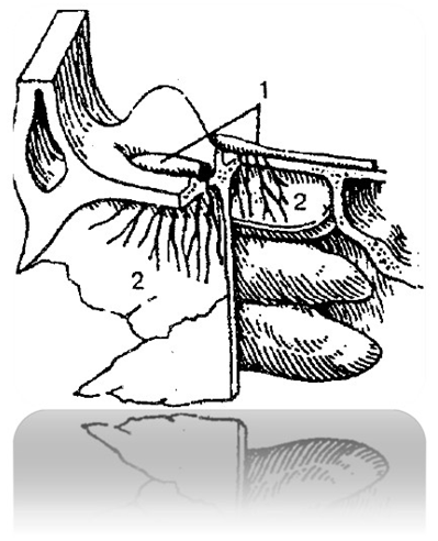
Fig. 23 Olfactory nerve 219 312 819 212
1 – olfactory bulbs 024 312 598 742
2 – olfactory nerves 219 312 819 212

Fig. 24 Optic nerve 448 817 918 217
1 – eyeball 519 321 819 288
2 – optic nerve 448 817 918 217
3 – orbital part 219 316 019 517
4 – intra-canalicular part 514 317 814 218
5 – intracranial part 219 321 919 821
6 – optic chiasm 010 216 319 517


Fig. 25 Oculomotor and trochlear nerves
819 321 919 821:
1 – crossing of the trochlear nerves 519 819 210 248
2 – trochlear nerve 498 064 318 712
3 – oculomotor nerve 519 217 519 217
4 – sympathetic root 548 571 918 221
5 – optic nerve (part) 448 817 918 217
6 – short ciliary nerves 478 521 928 321
7 – ciliary node 519 788 589 228
8 – inferior branch of the oculomotor nerve
219 888 999 617
9 – nasociliary root 519 512 879 002
10 – trigeminal nerve 549 319 818 711
11 – superior branch of the oculomotor nerve
501 048 998 118
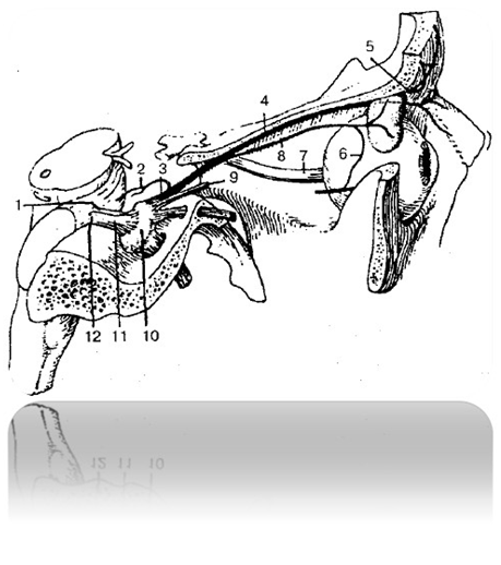
Fig. 26 Ophthalmic nerve (first branch of the trigeminal nerve) 914 815 914 715
1 – motor root 929 317 918 817
2 – tentorial (sheath) branch
929 317 918 513
3 – optic nerve 317 918 478 217
4 – frontal nerve 519 712 919 812
5 – supraorbital nerve 948 517 218 557
6 – communicating branch (with the zygomatic nerve)
514 827 918 527
7 – optic nerve 448 817 918 217
8 – lacrimal nerve 478 217 378 217
9 – nasociliary nerve 519 318 219 298
10 – trigeminal ganglion 418 217 218 217
11 – trigeminal nerve 549 319 818 711
12 – sensitive root 514 321 814 721
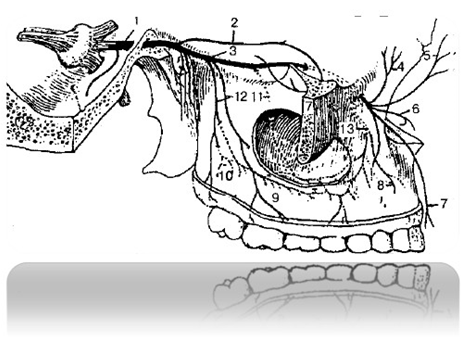
Fig. 27 Maxillary nerve (second branch
trigeminal nerve) 519 712 819 222:
1 – maxillary nerve 519 712 919 222
2 – zygomatic nerve 498 517 018 917
3 – infraorbital nerve 599 864 728 216
4 – lower branches of the eyelids 519 231 919 811
5 – external nasal branches 519 817 919 227
6 – internal nasal branches 498 515 918 225
7 – superior labial branches 514 273 518 223
8 – upper dental branches 519 816 319 777
9 – upper gingival branches 418 217 918 227
10 – upper dental plexus 519 621 978 911
11 – middle superior alveolar branch
514 371 948 211
12 – posterior superior alveolar branches
514 327 549 247
13 – anterior superior alveolar branches
548 217 217 319

Fig. 28 Mandibular nerve (third branch
trigeminal nerve) 514 321 814 221
1 – mandibular nerve 514 321 814 221
2 – lateral pterygoid nerve 214 712 814 212
3 – medial pterygoid nerve 519 712 819 212
4 – buccal nerve 489 061 278 671
5 – masseteric nerve 518 217 918 227
6 – auriculotemporal nerve 319 721 919 221
7 – anterior auricular nerves 248 655 448 755
8 – superficial temporal branches 429 321 899 411
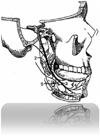
Fig. 29 Lingual nerve 214 318 714 818
1 – lingual nerve 214 318 714 818
2 – mylohyoid nerve 519 312 419 712
3 – lingual branches 819 418 519 718
4 – hypoglossal nerve 548 321 555 678
5 – submandibular parasympathetic ganglion
314 815 214 915
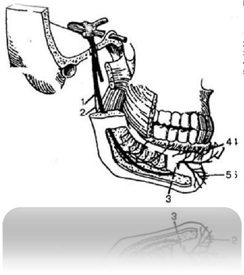
Fig. 30 Inferior alveolar nerve
891 318 519 458
1 – inferior alveolar nerve 891 318 519 458
2 – mylohyoid nerve 519 312 419 712
3 – mental nerve 319 814 819 914
4 – labial and gingival branches 514 813 914 608
5 – mental branches 319 812 819 312
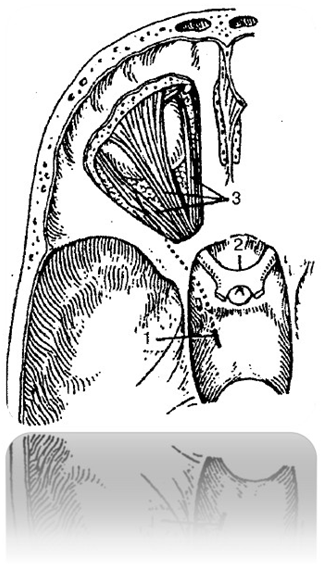
Fig. 31 Abducens nerve 514 517 214 812
1 – abducens nerve 514 517 214 812
2 – optic nerve 448 817 918 217
3 – eye muscles 548 712 818 912
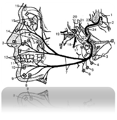
Fig. 32 Facial nerve 999 811 319 211:
1 – bottom of the IV ventricle 519 318 010 624
2 – nucleus of the facial nerve 668 317 918 517
3 – stylomastoid foramen 514 318 214 818
4 – branch to the posterior auricular muscle 919 312 819 512
5 – branch to the posterior belly of the digastric muscle 519 318 918 312
6 – branch to the stylohyoid muscle 514 812 819 412
7 – branches of the facial nerve to the facial muscles and subcutaneous
neck muscle 514 317 418 219
8 – branch to the muscle that depresses the angle of the mouth 514 317 214 317
9 – branch to the mentalis muscle 919 512 418 712
10 – branch to the muscle that lowers the lower lip 519 312 518 712
11 – branch to the buccinator muscle 418 317 814 217
12 – branch to the orbicularis oris muscle 219 379 891 472
13 – branch to the muscle that raises the upper labium 519 314 718 213
14 – branch to the zygomatic muscle 521 378 421 278
15 – branches to the orbicularis oculi muscle 498 781 398 217
16 – branches to the frontal belly of the epicranial muscle 548 371 898 217
17 – Drum string 519 712 891 421
18 – lingual nerve 214 318 714 818
19 – pterygopalatine ganglion 548 317 814 312
20 – trigeminal ganglion 418 217 218 217
21 – internal carotid artery 549 712 810 248
22 – intermediate nerve 219 381 648 719
23 – facial nerve 999 811 319 211
24 – vestibulocochlear nerve 548 217 918 421
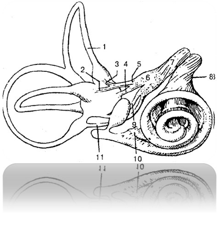
Fig. 33 Vestibular cochlear nerve 548 217 918 421:
1 – semicircular canals 841 918 219 312
2 – lateral ampullary nerve 519 471 898 371
3 – anterior ampullary nerve 548 317 918 221
4 – elliptical saccular nerve 519 328 499 228
5 – elliptical-saccular-ampullary nerve
514 312 814 882
6 – pre-door unit 719 317 919 817
7 – vestibular nerve 519 481 919 371
8 – cochlear nerve 518 317 918 221
9 – spherical saccular nerve 518 472 918 222
10 – cochlear ganglion (spiral ganglion of the cochlea)
914 712 814 212
11 – posterior ampullary nerve 219 473 218 223
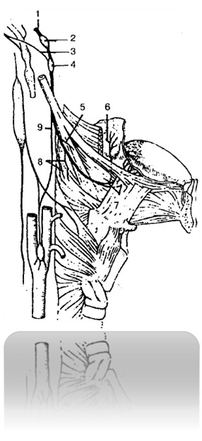
Fig. 34 Glossopharyngeal nerve 519 371 214 572:
1 – glossopharyngeal nerve 519 371 214 572
2 – upper node 501 784 968 781
3 – connecting branch 519 317 068 007
4 – lower node 319 216 519 428
5 – branch of the stylopharyngeus muscle 542 718 212 328
6 – almond branches 549 317 229 327
7 – lingual branches 819 418 519 718
8 – pharyngeal branches 498 217 228 417
9 – branch of the carotid sinus 319 421 219 221
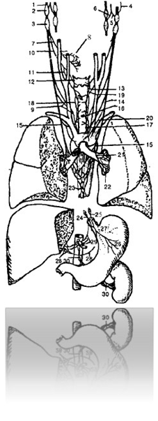
Fig. 35 Vagus nerve 489 981 728 221:
1 – vagus nerve 489 981 728 221
2 – upper node 501 784 968 781
3 – lower node 319 216 519 428
4 – meningeal branch 548 640 294 371
5 – auricular branch 519 318 919 287
6 – connecting branch 519 321 919 811
7 – pharyngeal branches 498 217 228 417
8 – pharyngeal plexus 519 315 919 885
9 – superior cervical cardiac branches 498 712 319 882
10 – superior laryngeal nerve 498 882 319 982
11 – external branch 519 831 918 281
12 – internal branch 498 215 298 195
13 – communicating branch with the recurrent laryngeal nerve 591 061 974 891
14 – lower cervical cardiac branches 498 761 998 251
15 – recurrent laryngeal nerve 518 472 888 912
16 – tracheal branches 919 810 499 310
17 – esophageal branches 519 512 319 812
18 – inferior laryngeal nerve 514 312 814 222
19 – communicating branch with the internal laryngeal branch 519 321 718 221
20 – thoracic cardiac branches 989 312 918 212
21 – bronchial branches 619 321 819 221
22 – pulmonary plexus 428 317 888 917
23 – esophageal plexus 219 641 298 581
24 – front wandering trunk 214 217 914 817
25 – rear wandering trunk 219 317 219 228
26 – anterior gastric branches 289 317 299 277
27 – posterior gastric branches 298 312 678 212
28 – hepatic branches 214 213 219 312
29 – celiac branches 298 012 718 202
30 – renal branches 219 317 209 717
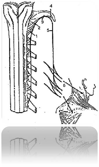
Fig. 36 Accessory nerve 489 917 319 712:
1 – spinal roots 519 641 918 471
2 – cranial roots (vagal part) 548 217 319 227
3 – trunk of the accessory nerve 419 061 918 432
4 – internal branch 498 215 298 195
5 – external branch 519 831 918 281
6 – muscular branches 214 312 814 282
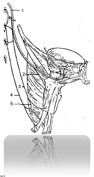
Fig. 37 Hypoglossal nerve and cervical
(sublingual) loop 214 332 817 728:
1 – hypoglossal nerve 548 321 555 678
2 – thyrohyoid branch 519 513 419 213
3 – front spine 519 317 819 228
4 – back root 409 272 819 322
5 – cervical (sublingual) loop 841 332 817 798
6 – lingual branches 819 418 519 718
Spinal nerves 489 218 918 217

Fig. 38 Spinal nerve 489 218 918 217:
1 – spinal nerve trunk 848 312 888 202
2 – anterior (motor) root 248 317 881 204
3 – posterior (sensory) root 512 312 489 202
4 – root threads 514 327 988 189
5 – spinal (sensory) node
918 312 889 202
6 – medial part of the posterior branch 819 317 228 227
7 – lateral part of the posterior branch 519 312 819 272
8 – posterior branch 548 715 888 215
9 – anterior branch 598 671 098 317
10 – white branch 519 064 319 711
11 – gray branch 229 829 318 912
12 – meningeal branch 498 517 328 777
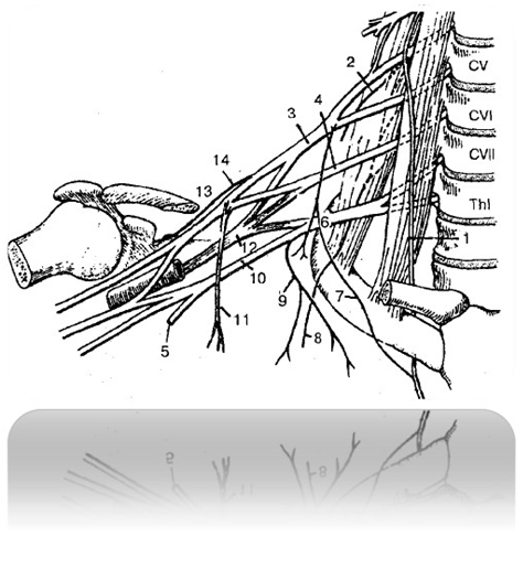
Fig. 39 Brachial plexus 312 314 512 214:
1 – phrenic nerve 519 378 219 888
2 – dorsal nerve of the scapula 888 888 919 017
3 – upper trunk of the brachial plexus 428 217 228 277
4 – middle trunk of the brachial plexus 514 312 814 222
5 – subclavian trunk 498 217 289 277
6 – lower trunk of the brachial plexus 748 213 888 213
7 – accessory phrenic nerves 067 214 327 224
8 – long thoracic nerve 519 641 598 714
9 – medial thoracic nerve 418 722 888 222
10 – lateral thoracic nerve 214 317 914 777
11 – medial bundle 819 312 819 314
12 – rear bundle 889 212 478 312
13 – lateral bundle 514 312 889 212
14 – suprascapular nerve 418 319 218 912

Fig. 40 Nerves of the shoulder, forearm and hand 412 818 219 314:
A - nerves of the shoulder 429 561 989 079:
1 - medial cutaneous nerve of the arm and medial cutaneous nerve
forearms 428 312 518 212
2 – median nerve 498 717 818 217
3 – brachial artery 598 641 389 071
4 – ulnar nerve 319 817 919 016
5 – biceps brachii (distal end) 598 064 389 517
6 – radial nerve 514 317 814 212
7 – shoulder muscle 319 717 819 317
8 – musculocutaneous nerve 512 314 212 814
9 – biceps brachii (proximal end)
514 319 064 818
B – nerves of the forearm and hand 218 001 209 317:
1 – median nerve 498 717 818 217
2 – pronator teres (crossed) 214 888 219 317
3 – ulnar nerve 319 817 919 016
4 – deep flexor of the fingers 213 814 818 217
5 – anterior interosseous nerve 548 312 819 272
6 – dorsal branch of the ulnar nerve 428 577 928 227
7 – deep branch of the ulnar nerve 458 317 218 757
8 – superficial branch of the ulnar nerve 419 312 819 422
9 – pronator quadratus (crossed) 489 016 718 941
10 – superficial branch of the radial nerve 548 217 328 227
11 – brachioradialis muscle (crossed) 429 318 229 718
12 – radial nerve 514 317 814 212
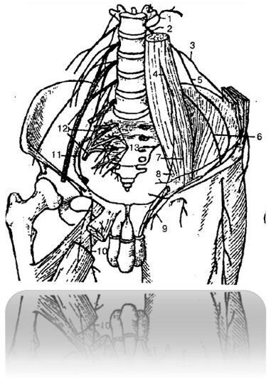
Fig. 41 Lumbosacral plexus 214 712 814 328:
1 – posterior branches of the lumbar nerves 418 712 818 322
2 – anterior branches of the lumbar nerves 514 372 814 222
3 – iliohypogastric nerve 514 371 814 211
4 – genitofemoral nerve 518 317 989 316
5 – ilioinguinal nerve 518 471 299 317
6 – lateral cutaneous nerve of the thigh 519 618 919 818
7 – femoral branch 514 317 814 818
8 – sexual branch 528 712 328 912
9 – anterior scrotal nerves 428 319 718 219
10 – anterior branch of the obturator nerve 514 718 219 317
11 – obturator nerve 589 317 919 217
12 – lumbosacral plexus 214 712 814 328
13 – anterior branches of the sacral plexus 548 312 219 228
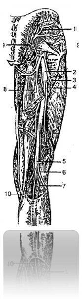
Fig. 42 Nerves of the gluteal region and posterior surface
hips 214 718 219 312:
1 – superior gluteal nerve 419 715 819 215
2 – sciatic nerve 898 919 719 828
3,4 – muscular branches of the sciatic nerve 214 312 814 712
5 – tibial nerve 518 714 818 914
6 – common peroneal nerve 498 217 998 717
7 – lateral cutaneous nerve of the calf 492 517 212 817
8 – posterior cutaneous nerve of the thigh 514 317 218 277
9 – inferior gluteal nerve 819 215 518 225
10 – medial dorsal cutaneous nerve 514 312 814 872
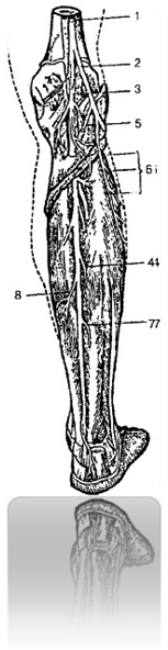
Fig. 43 Nerves of the leg (posterior surface) 548 217 218 887:
1 – sciatic nerve 898 919 719 828
2 – common peroneal nerve 498 217 998 717
3 – tibial nerve 514 812 919 222
4, 7, 8 – muscular branches of the tibial nerve 548 714 818 214
5 – lateral cutaneous nerve of the calf 492 517 212 817
6 – muscular branches of the peroneal nerve 548 219 319 878
VEGETATIVE (AUTONOMIC) NERVOUS SYSTEM
514 312 819 981
Sympathetic part of the autonomic nervous system 891 418 318 888
Parasympathetic part of the autonomic nervous system 418 217 318 918
Cytoarchitectonic fields of the cerebral cortex 219 047 819 215
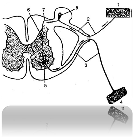
Rice. 44 Reflex arc 219 317 819 892:
1 - nerve endings of a sensory neuron in the skin
428 317 918 217
2 - peripheral process of a sensory neuron
591 489 766 281
3 – axon of motor cell 428 312 918 212
4 – nerve ending in muscle 519 317 918 287
5 – motor cell of the anterior horn 491 218 519 328
6 – interneuron 314 517 214 817
7 – central process of the sensory neuron
512 216 219 327
8 – spinal ganglion 428 312 219 217
Signaling systems 891 312 918 412
SENSE ORGANS 214 712 514 312
Organ of vision 219 317 989 312
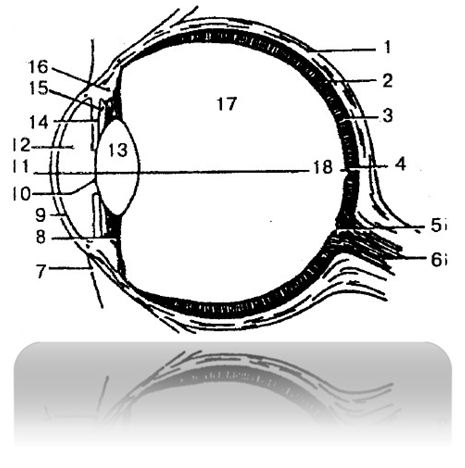
Fig. 45 Eye 514 317 814 917
1 – sclera 928 317 818 917
2 – choroid 218 712 819 312
3 – retina 319 218 918 217
4 – central fossa 548 614 719 641
5 – Blind Spot 489,000 909,216
6 – optic nerve 448 817 918 217
7 – conjunctiva 528 317 312 819
8 – ciliary ligament 898 312 519 482
9 – cornea 489 317 219 217
10 – pupil 489 319 218 213
11, 18 – optical axis 519 317 419 817
12 – front camera 489 715 219 816
13 – lens 519 321 819 221
14 – iris 428 218 318 219
15 – rear camera 489 312 918 216
16 – ciliary muscle 219 312 719 312
17 – vitreous body 519 322 819 212
Accessory organs of the eye 219 899 319 214
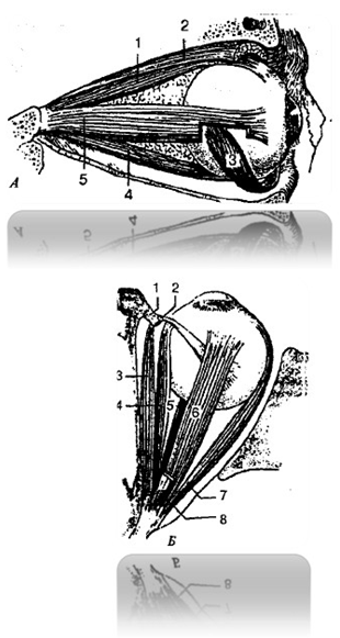
Fig. 46 Muscles of the eyeball 512 901 318 201:
A – lateral view:
1 – superior rectus muscle 498 617 219 190
2 – muscle that lifts the upper eyelid 512 419 312 228
3 – inferior oblique muscle 319 618 204 881
4 – inferior rectus muscle 219 317 219 827
5 – lateral rectus muscle 328 421 898 712
B – top view:
1 – block 229 457 298 788
2 – sheath of the tendon of the superior oblique muscle
248 272 458 299
3 – superior oblique muscle 219 312 919 802
4 – medial rectus muscle 898 782 988 312
5 – inferior rectus muscle 219 317 219 827
6 – superior rectus muscle 498 617 219 190
7 – lateral rectus muscle 328 421 898 712
8 – muscle that lifts the upper eyelid 512 419 312 228
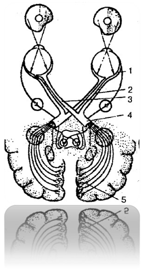
Fig. 47 Visual analyzer 219 318 719 817
1 – retina 319 218 918 217
2 – uncrossed fibers of the optic nerve 214 317 819 007
3 – crossed fibers of the optic nerve 719 300 800 111
4 – optic tract 519 218 919 245
5 – cortical analyzer 519 312 819 227
Organ of hearing and balance 248 712 318 222
Fig. 48 Vestibule-cochlear organ (organ
hearing and balance) 248 712 318 222:
1 – superior semicircular canal 498 312 818 221
2 – vestibule 948 218 298 820
3 – snail 321 018 204 516
4 – auditory nerve 458 912 312 819
5 – carotid artery 428 713 828 213
6 – auditory tube 429 318 919 228
7 – tympanic cavity 519 317 919 827
8 – eardrum 429 317 229 817
9 – external auditory canal 519 421 919 811
10 – external auditory canal 521 917 319 817
11 – auricle 421 918 518 717
12 – hammer 521 328 421 891
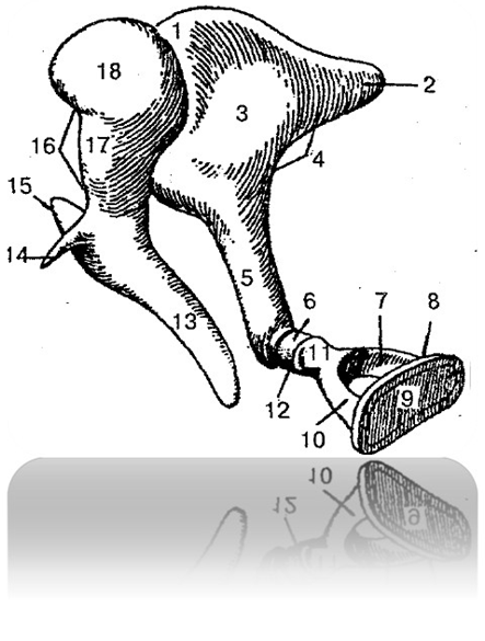
Fig. 49 Auditory ossicles 428 317 218 227
1 – incudomalleolar joint 214 312 914 212
2 – short anvil leg 512 612 218 377
3 – anvil body 889 891 892 712
4 – anvil 521 488 711 918
5 – long anvil leg 548 712 528 312
6 – lenticular process 528 219 312 219
7 – rear leg of the stirrup 428 517 928 717
8 – stirrup 498 714 889 216
9 – stirrup base 378 819 498 881
10 – anterior stirrup leg 488 711 294 301
11 – stirrup head 481 499 816 701
12 – incudostapedial joint 797 014 398 481
13 – hammer handle 878 421 891 216
14 – anterior process of malleus 219 054 398 716
15 – lateral process of malleus 481 898 714 849
16 – hammer 521 328 421 891
17 – neck of malleus 891 219 311 919
18 – hammer head 214 312 814 918
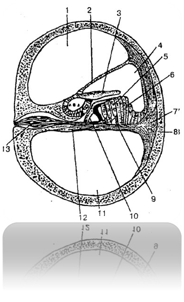
Fig. 50 Cochlear canal (transverse view):
1 – vestibule staircase 219 317 918 516
2 – vestibular wall of the cochlear duct 498 641 919 319
3 – cover membrane 498 516 679 210
4 – cochlear duct 312 814 219 322
5 – auditory cells with cilia 512 418 622 898
6 – support cells 548 312 988 822
7 – spiral ridge (spiral ligament) 528 318 618 227
8 – cochlear bone tissue 428 219 319 891
9 – support cage 294 316 819 511
10 – Corti cells-pillars 214 316 719 816
11 – drum ladder 918 421 519 317
12 – basilar plate 514 321 898 218
13 – nerve cells of the spiral ganglion 428 317 428 527
Taste organ 419 317 819 227
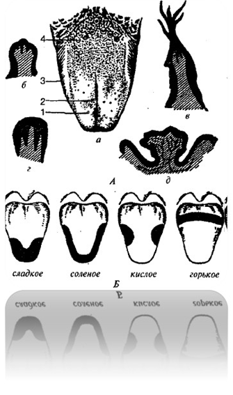
Fig. 51 Papillae of the tongue. Taste fields of the tongue 419 381 489 294:
A – tongue papillae 419 381 489 294
a - general view 914 819 319 821
b – fungiform papilla 314 218 914 888
in - filiform papilla 589 617 298 471
g - foliate papilla 418 644 319 515
d - circumvallate papilla 891 319 481 617
1 – fungiform papillae 314 218 914 888
2 – filiform papillae 589 617 298 471
3 – foliate papillae 418 644 319 515
4 – circumvallate papillae 891 319 481 617
B – taste fields of the tongue 419 317 819 217
Olfactory organ 914 782 214 389
Leather 519 606 901 319
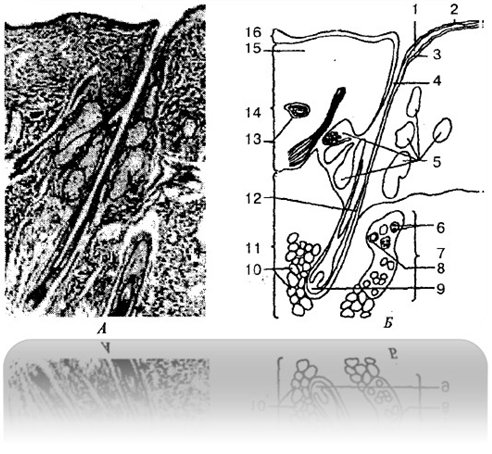
Fig. 52 Human skin: (A – transverse view,
B - visible structures) 519 606 901 319
1 – granular layer 514 317 814 297
2 – stratum corneum 548 671 918 311
3 – Malpighian layer 914 917 414 897
4 – hair follicle 314 912 814 889
5 – sebaceous glands 498 791 229 321
6 – secretory cells 519 818 229 398
7 – sweat gland 519 488 598 719
8 – gland duct 891 098 789 016
9 – hair papilla 918 781 298 391
10 – subcutaneous fat cells 898 712 918 312
11 – subcutaneous tissue 519 861 719 211
12 – hair shaft part 598 061 214 711
13 – blood vessel 217 918 294 888
14 – derma 498 718 519 317
15 – elastic and collagen fibers
519 618 718 215
16 – epidermis 598 718 889 888
Numerical concentrations on the skin activate the restoration of organ matter in connection with the entire organism. The division of the internal space of the organism and the external space in relation to the organism by the skin allows numerical concentrations on the skin to control events in the organism and outside it more widely than numerical concentrations on other matter.
Therefore, when managing events as an accelerator
element can be applied to the skin in concentrations. Micro and macro objects harmful to the body, including infections
and the like, you can remove them from the body faster or prevent them from infecting the body using
Numerical concentrations on the skin.
BONES 214 318 214 818
BONE CONNECTIONS 814 312 214 712
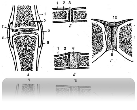
Fig. 53 Types of bone connections:
A – joint 314 812 514 212
B - fibrous connection 719 312 819 212
B – synchondrosis (cartilaginous joint) 314 217 914 818
G – symphysis 519 488 617 314
1 – periosteum 598 061 318 041
2 – bone 418 517 918 217
3 – fibrous connective tissue 519 318 219 417
4 – cartilage 314 217 914 819
5 – synovial membrane 214 317 914 817
6 – fibrous membrane 319 217 916 074
7 – articular cartilage 314 217 914 914
8 – joint cavity 312 817 918 217
9 – interpubic disc space 514 318 217 218
10 – interpubic disc 219 312 219 212
Skeleton of the trunk 219 314 819 217
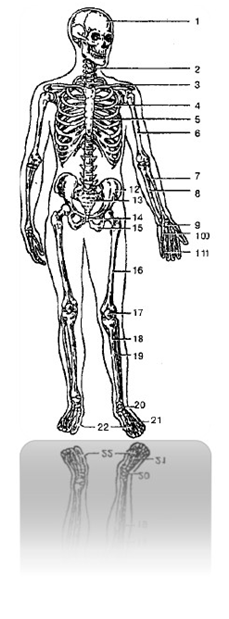
Fig. 54 Human skeleton 219 317 918 428:
1 – skull 519 515 919 894
2 – spinal column 328 488 984 012
3 – collarbone 409 382 984 712
4 – rib 519 312 918 722
5 – sternum 518 417 289 897
6 – humerus 524 377 914 877
7 – radius 529 321 919 742
8 – ulna 529 341 419 811
9 – carpal bones 318 712 918 212
10 – metacarpal bones 214 371 814 911
11 – phalanges of fingers 548 317 918 217
12 – ilium 218 317 228 917
13 – sacrum 514 716 814 226
14 – pubic bone 512 478 212 238
15 – ischium 498 216 748 227
16 – femur 918 275 784 325
17 – patella 421 891 529 328
18 – tibia 918 321 989 711
19 – fibula 498 217 888 917
20 – tarsal bones 498 215 298 315
21 – metatarsal bones 494 216 894 317
22 – phalanges of the toes 548 321 984 671
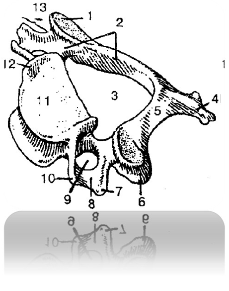
Fig. 55 Cervical vertebra 219 213 319 721
1 – superior articular process 519 317 819 217
2 – vertebral arch 519 312 919 212
3 – vertebral foramen 489 016 219 715
4 – spinous process 548 312 848 212
5 – vertebral arch plate 517 218 317 918
6 – inferior articular process 517 919 217 398
7 – posterior tubercle 498 641 518 919
8 – groove of spinal nerve 219 317 819 218
9 – opening of the transverse process 514 318 218 214
10 – anterior tubercle 519 218 218 914
11 – vertebral body 317 689 318 918
12 – body hook 316 219 819 478
13 – transverse process 548 610 989 241
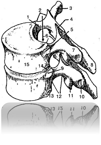
Fig. 56 Thoracic vertebrae 498 614 319 712:
1 – pedicle of the vertebral arch 498 317 218 217
2 – superior vertebral notch 598 641 398 011
3, 7 – transverse process 598 642 319 811
4 – superior articular process 219 715 319 215
5, 9 – superior costal fossa 549 312 814 212
6 – spinal canal 521 314 818 214
8 – spinous process 518 617 218 141
10 – costal fossa of the transverse process 821 319 921 819
11 – inferior articular process 528 644 328 016
12 – lower vertebral notch 512 314 812 214
13, 14 – lower costal fossa 019 712 219 312
15 – vertebral body 517 219 319 617
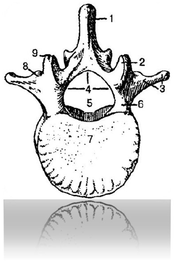
Fig. 57 Lumbar vertebra (top view) 519 317 819 218:
1 – spinous process 513 219 813 919
2 – superior articular process 519 617 299 017
3 – costal process 317 814 214 917
4 – vertebral arch 391 498 016 217
5 – vertebral foramen 828 317 918 217
6 – pedicle of the vertebral arch 498 317 218 227
7 – vertebral body 598 641 319 071
8 – additional process 518 431 219 917
9 – mastoid process 519 817 919 217
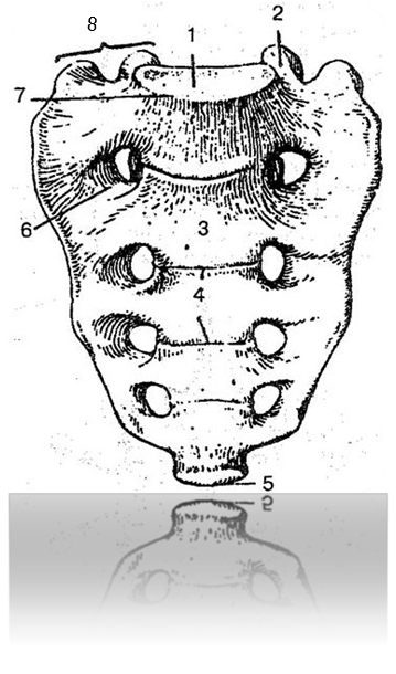
Fig. 58 Sacrum (front view) 514 716 814 226:
1 – base of the sacrum 519 614 319 812
2 – superior articular process 519 328 919 228
3 – anterior surface of the sacrum 549 218 319 228
4 – transverse lines 428 213 328 333
5 – apex of the sacrum 408 217 229 327
6 – anterior sacral openings 489 213 217 289
7 – Cape 428 327 828 227
8 – lateral part 319 712 919 212
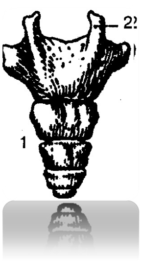
Fig. 59 Coccyx (rear view) 519 513 819 213:
1 – coccyx 519 513 819 213
2 – coccygeal horn 428 617 988 064
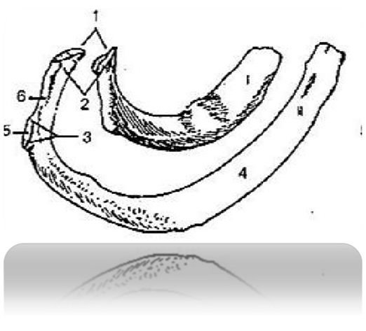
Fig. 60 First and second ribs (top view)
214 712 814 312:
1 – articular surface of the head of the rib
512 318 912 218
2 – rib head 214 718 218 214
3 – tubercle of the rib 518 380 218 910
4 – rib body 538 712 818 222
5 – articular surface of the tubercle of the cost
419 710 819 210
6 – neck of rib 498 217 218 317
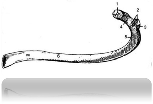
Fig. 61 Seventh rib (internal
surface) 519 318 919 218:
1 – articular surface of the head of the rib
598 641 398 061
2 – articular surface of the tubercle of the cost
489 681 717 414
3 – tubercle of the rib 518 714 318 214
4 – neck of rib 074 216 519 421
5 – rib angle 219 718 218 312
6 – rib body 217 419 218 219
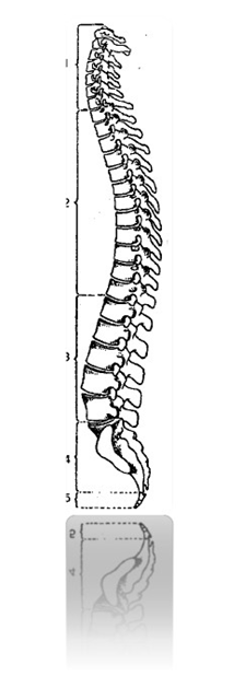
Fig. 62 Vertebral column 214 217 000 819:
1 – cervical vertebrae 312 218 212 918
2 – thoracic vertebrae 498 614 319 712
3 – lumbar vertebrae 498 217 218 227
4 – sacrum 514 716 814 226
5 – coccyx 519 513 819 213
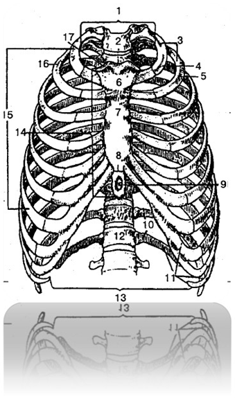
Fig. 63 Skeleton of the thorax (front view)
248 668 712 298:
1 – upper chest aperture 520 319 210 299
2 – jugular notch 548 717 818 997
3 – ribs (1 – 12) 431 898 211 328
4 – first rib 317 814 217 214
5, 16 – second rib 518 513 918 913
6 – sternum handle 819 312 219 312
7 – body of the sternum 514 318 219 217
8 – articulation between the body of the sternum and the xiphoid
process 512 318 918 212
9 – xiphoid process 219 318 719 228
10 – oscillating ribs (11 – 12) 514 217 214 317
11 – false ribs (8 – 10) 548 212 228 312
12 – thoracic vertebra 542 317 212 227
13 – lower thoracic aperture 009 217 819 317
14 – sternum 518 417 289 897
15 – true ribs (1 – 7) 789 641 298 514
17 – clavicular notch 312 814 212 418
Skeleton of the head 231 138 918 212
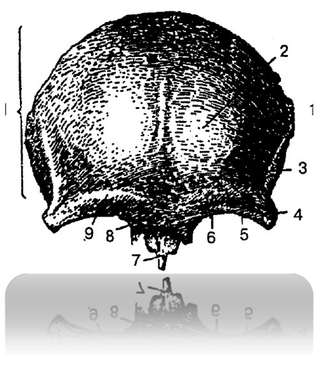
Fig. 64 Frontal bone 248 003 398 213:
1 – scales 214 317 818 217
2 – frontal tubercle 219 317 818 227
3 – temporal line 248 312 298 222
4 – zygomatic process 248 714 318 214
5 – supraorbital margin 249 312 289 228
6 – supraorbital foramen 312 278 229 312
7 – nose section 248 217 228 327
8 – glabella (glabella) 814 712 214 312
9 – brow ridge 428 517 228 917
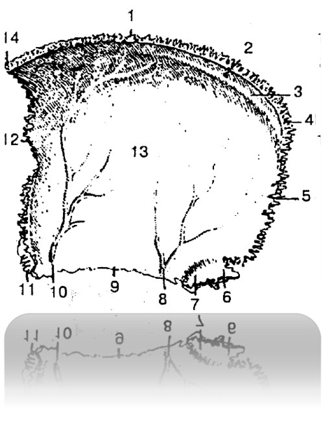
Fig. 65 Right parietal bone (internal
surface) 312 718 212 218
1 – sagittal edge 519 417 319 816
2 – parietal foramen 312 219 218 271
3 – groove of the superior sagittal sinus
214 317 228 271
4 – occipital angle 248 712 219 220
5 – occipital margin 319 714 219 514
6 – mastoid angle 314 217 214 227
7 – groove of the sigmoid sinus 278 213 228 913
8 – 10 – groove of the middle meningeal artery
514 317 814 717
11 – wedge angle 594 018 294 318
12 – frontal margin 364 618 972 318
13 – inner surface 234 712 814 212
14 – frontal angle 514 001 814 321
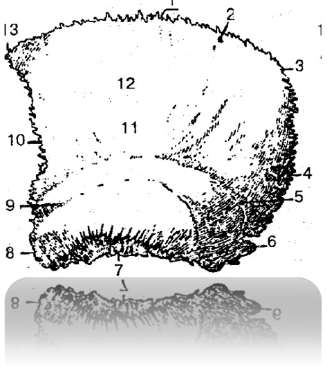
Fig. 66 Left parietal bone (external
surface) 326 718 216 718:
1 – sagittal edge 519 417 319 816
2 – parietal foramen 312 219 218 271
3 – occipital angle 248 712 219 220
4 – occipital margin 319 714 219 514
5 – upper temporal line 548 312 718 212
6 – mastoid angle 314 217 214 227
7 – scaly edge 298 714 888 914
8 – wedge angle 594 018 294 318
9 – lower temporal line 548 317 818 717
10 – frontal margin 364 618 972 318
11 – parietal tubercle 219 317 918 227
12 – outer surface 519 318 719 288
13 – frontal angle 514 001 814 321
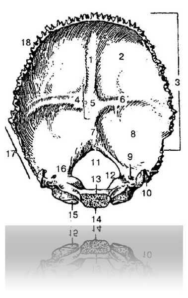
Fig. 67 Occipital bone (inner surface)
214 712 219 312:
1 – groove of the superior sagittal sinus 214 317 228 271
2 – medullary fossa 549 312 919 217
3 – occipital scales 598 317 919 217
4 – cross-shaped elevation 519 312 299 812
5 – internal occipital protuberance 312 818 712 918
6 – groove of the transverse sinus 594 361 298 061
7 – internal occipital crest 521 316 919 488
8 – cerebellar fossa 219 213 919 223
9 – condylar canal 231 918 298 221
10 – jugular process 498 317 998 227
11 – Large hole 201 398 721 778
12 – jugular tubercle 548 715 328 225
13 – basilar part 549 713 919 223
14 – pharyngeal tubercle 548 712 228 312
15 – occipital condyle 549 317 819 223
16 – lateral part 549 717 229 237
17 – mastoid margin 542 713 222 203
18 – lambdoid margin 519 321 009 811

Fig. 68 Occipital bone (posterior and inferior view)
548 717 218 317:
1 – occipital scales 598 317 919 217
2 – jugular tubercle 548 715 328 225
3 – slope 319 778 219 228
4 – large hole 201 398 721 778
5 – condylar canal 231 918 298 221
6 – occipital condyle 549 317 819 223
7 – condylar fossa 539 812 918 222
8 – lower nuchal line 514 701 814 321
9 – external occipital crest 514 312 814 722
10 – superior nuchal line 548 717 888 999
11 – highest nuchal line 599 000 089 319
12 – occipital platform 538 721 918 211
13 – external occipital protuberance 429 312 819 228
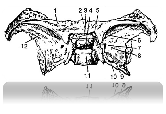
Fig. 69 Sphenoid bone (top view)
321 694 019 181:
1 – small wing (left) 329 717 229 317
2 – body 429 811 319 321
3 – pre-cross groove 314 717 814 217
4 – pituitary fossa 519 317 919 218
5 – visual channel 538 712 918 222
6 – superior orbital fissure 409 505 898 305
7 – round hole 514 318 618 711
8, 12 – large wings 217 318 917 228
9 – oval hole 298 714 318 214
10 – spinous foramen 219 542 319 712
11 – Saddle back 548 713 918 213
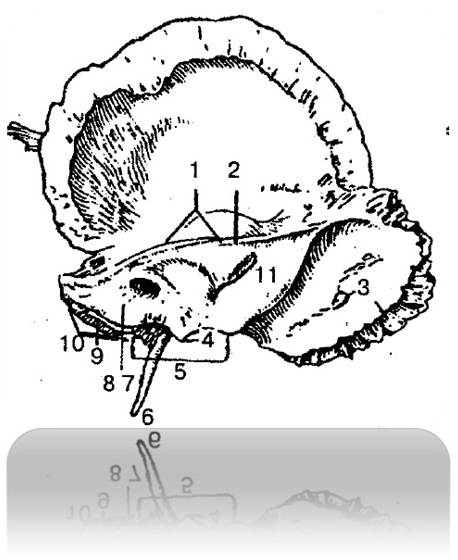
Fig. 70 Right temporal bone (internal
surface) 549 317 914 317:
1 – the upper edge of the pyramid 319 716 818 012
2 – groove of the superior petrosal sinus 549 312 819 229
3 – groove of the sigmoid sinus 278 213 228 913
4 – internal process 214 712 914 229
5 – jugular notch 531 918 911 218
6 – styloid process 598 312 818 212
7 – internal auditory canal 548 211 918 211
8 – internal auditory canal 531 988 411 888
9 – groove of the inferior petrosal sinus 219 213 919 733
10 – the back edge of the pyramid 214 313 219 733
11 – back surface of the pyramid 519 312 814 222
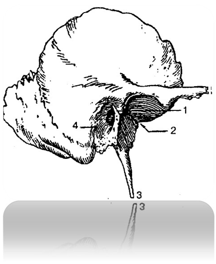
Fig. 71 Right temporal bone (external
surface) 519 312 419 812:
1 – tympanic-scaly gap 514 317 814 317
2 – petrotympanic fissure 548 321 948 221
3 – styloid process 598 312 818 212
4 – tympanomastoid fissure 598 712 898 223

Bones of the facial part of the skull 219 715 819 815
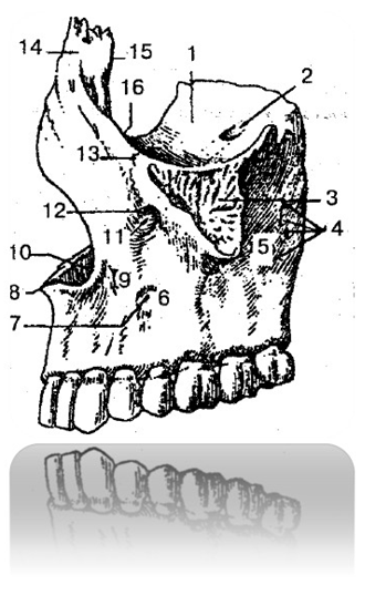
Fig. 72 Upper jaw (lateral view)
parties) 519 371 919 811:
1 – orbital surface 398 216 718 226
2 – infraorbital groove 319 717 819 227
3 – zygomatic process 419 312 214 222
4 – alveolar openings 214 712 814 229
5 – temporal surface 538 722 918 222
6 – front surface 548 888 019 648
7 – canine fossa 539 717 819 317
8 – anterior nasal spine 529 513 919 813
9 – body of the upper jaw 548712 818 212
10 – nasal notch 548 716 298 444
11 – infraorbital canal 319 717 819 217
12 – infraorbital foramen 489 061 298 541
13 – zygomaticomaxillary suture 214 711 898 211
14 – frontal process 590 421 019 481
15 – lacrimal edge 548 884 918 888
16 – infraorbital margin 512 219 312 919

Fig. 73 Left upper jaw (medial view)
parties) 421 718 911 328
1 – frontal process 590 421 019 481
2 – nasal surface 598 648 319 711
3 – anterior nasal spine 529 513 919 813
4 – pterygopalatine groove 428 321 814 221
5 – maxillary (maxillary) sinus 519 321 814 471
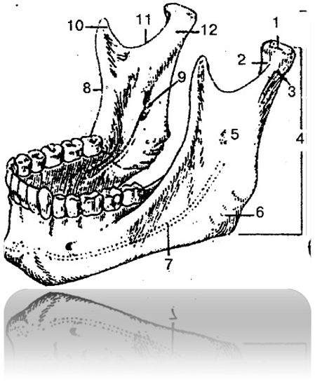
Fig. 74 Lower jaw 514 712 814 312:
1 – head of the lower jaw 548 321 848 721
2 – pterygoid fossa 519 317 919 007
3 – neck of the lower jaw 319 814 919 714
4, 5 – branches of the lower jaw 518 317 918 001
6 – angle of the lower jaw 548 219 289 008
7 – lower jaw canal 009 217 319 227
8 – temporal crest 418 317 228 227
9 – opening of the lower jaw 489 201 319 871
10 – coronoid process 528 317 918 228
11 – lower jaw notch 419 317 819 828
12 – condylar process 891 319 898 789
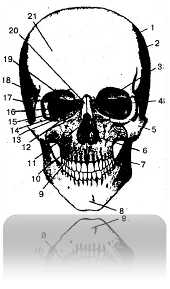
Fig. 75 Human skull (front view) 829 317 229 817:
1 – coronal suture 419 289 917 814
2 – parietal bone 519 987 219 317
3 – orbital part of the frontal bone 998 261 378 471
4 – orbital surface of the greater wing of the sphenoid bone
548 261 378 213
5 – zygomatic bone 899 817 818 317
6 – inferior nasal concha 478 218 918 217
7 – upper jaw 521 718 221 918
8 – Mental protrusion of the lower jaw 518 001 219 671
9 – nasal cavity 428 317 818 227
10 – coulter 429 317 819 228
11 – perpendicular plate of the ethmoid bone
501 608 919 219
12 – orbital surface of the maxilla 428 712 818 212
13 – inferior orbital fissure 429 731 819 221
14 – lacrimal bone 209 605 319 205
15 – orbital plate of the ethmoid bone 398 667 818 917
16 – superior orbital fissure 409 505 898 305
17 – squamous part of the temporal bone 428 319 288 299
18 – zygomatic process of the frontal bone 248 714 318 214
19 – opening of the visual canal 498 712 818 222
20 – nasal bone 518 314 818 214
21 – frontal tubercle 219 317 818 227
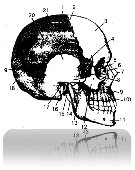
Fig. 76 Human skull (side view) 829 317 229 817:
1 – parietal bone 519 987 219 317
2 – coronal suture 419 289 917 814
3 – frontal tubercle 219 317 818 227
4 – temporal surface of the greater wing of the sphenoid
bones 514 317 049 612
5 – orbital plate of the ethmoid bone 398 667 818 917
6 – lacrimal bone 209 605 319 205
7 – nasal bone 518 314 818 214
8 – temporal fossa 548 312 448 212
9 – anterior nasal spine 529 513 919 813
10 – body of the upper jaw 518 312 818 212
11 – lower jaw 514 712 814 312
12 – zygomatic bone 899 817 818 317
13 – zygomatic arch 528 317 918 917
14 – styloid process 598 312 818 212
15 – condylar process of the mandible 891 319 898 789
16 – mastoid process 918 217 319 817
17 – external auditory canal 519 421 919 811
18 – lambdoid suture 428 711 318 911
19 – squama of the occipital bone 598 317 919 217
20 – upper temporal line 548 312 718 212
21 – squamous part of the temporal bone 428 319 288 299
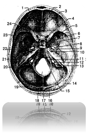
Fig. 77 Internal base of the skull 519 318 418 917:
1 – orbital part of the frontal bone 998 261 378 471
2 – cockscomb 428 312 818 222
3 – lattice plate 428 217 328 917
4 – visual channel 538 712 918 222
5 – pituitary fossa 519 317 919 218
6 – saddle back 513 988 813 488
7 – round hole 514 318 618 711
8 – oval hole 298 714 318 214
9 - Torn hole 529 317 216 489
10 – spinous foramen 219 542 319 712
11 – internal auditory canal 548 211 918 211
12 – jugular foramen 589 317 919 897
13 – sublingual canal 319 317 919 777
14 – lambdoid suture 428 711 318 911
15 – slope 319 778 219 228
16 – groove of the transverse sinus 594 361 298 061
17 – internal occipital protuberance 312 818 712 918
18 – foramen magnum 549 317 819 227
19 – occipital scales 598 317 919 217
20 – groove of the sigmoid sinus 278 213 228 913
21 – pyramid (petrous part) of the temporal bone
498 317 014 219
22 – squamous part of the temporal bone 428 319 288 299
23 – greater wing of the sphenoid bone 222 719 333 419
24 – lesser wing of the sphenoid bone 213 914 817 977
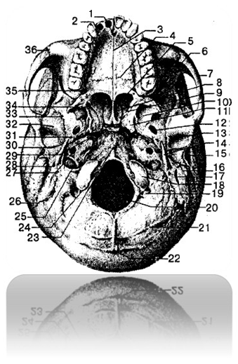
Fig. 78 External base of the skull 894 312 918 322:
1 – palatine process of the maxilla 819 312 419 222
2 – incisal opening 498 555 978 377
3 – median palatine suture 519 312 919 222
4 – transverse palatine suture 478 217 378 227
5 – choana 529 371 999 811
6 – inferior orbital fissure 429 731 819 221
7 – zygomatic arch 528 317 918 917
8 – wing of coulter 918 312 818 212
9 – pterygoid fossa 519 317 919 007
10 – lateral plate of the pterygoid process
548 321 918 221
11 – pterygoid process 219 311 919 211
12 – oval hole 298 714 318 214
13 – mandibular fossa 558 912 918 222
14 – styloid process 598 312 818 212
15 – external auditory canal 519 421 919 811
16 – mastoid process 918 217 319 817
17 – mastoid notch 519 312 819 221
18 – occipital condyle 549 317 819 223
19 – condylar fossa 539 812 918 222
20 – foramen magnum 549 317 819 227
21 – lower nuchal line 514 701 814 321
22 – external occipital protuberance 429 312 819 228
23 – pharyngeal tubercle 548 712 228 312
24 – condylar canal 231 918 298 221
25 – jugular foramen 589 317 919 897
26 – occipito-mastoid suture 214 312 827 488
27 – external carotid orifice 389 219 217 419
28 – stylomastoid foramen 514 318 214 818
29 - Torn hole 529 317 216 489
30 – petrotympanic fissure 548 321 948 221
31 – spinous foramen 219 542 319 712
32 – articular tubercle 288 412 298 322
33 – wedge-scale suture 298 717 298 277
34 – wing hook 598 328 219 830
35 – greater palatine foramen 219 498 817 312
36 – zygomaticomaxillary suture 214 711 898 211
Skeleton of limbs 548 212 788 272
Bones of the upper limb 971 981 319 212
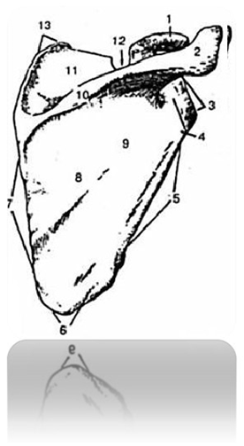
Fig. 79 Right shoulder blade (posterior view) 429 312 819 312:
1 – coracoid process 918 318 519 714
2 – acromion 529 319 919 712
3 – lateral angle 498 712 519 282
4 – neck of the scapula 498 710 218 220
5 – lateral edge 489 770 919 220
6 – lower corner 319 814 919 814
7 – medial edge 519 311 819 911
8 – back surface of the scapula 918 712 514 317
9 – infraspinous fossa 219 712 819 222
10 – scapula spine 498 712 328 822
11 – supraspinatus fossa 512 488 912 988
12 – shoulder blade cut 319 714 819 214
13 – top corner 489 617 319 441
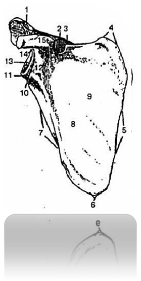
Fig. 80 Right shoulder blade (front view) 429 312 819 312:
1 – articular surface of acromion 519 311 919 211
2 – shoulder blade cut 319 714 819 214
3 – scapula spine 498 712 328 822
4 – top corner 489 617 319 441
5 – medial edge 519 311 819 911
6 – lower corner 319 814 919 814
7 – lateral edge 489 770 919 220
8 – subscapular fossa 528 621 389 710
9 – costal surface 519 412 819 312
10 – neck of the scapula 498 710 218 220
11 – subarticular tubercle 212 317 412 917
12 – lateral corner 528 318 914 818
13 – medial edge 548 912 928 312
14 – supra-articular tubercle 488 312 818 222
15 – coracoid process 918 318 519 714
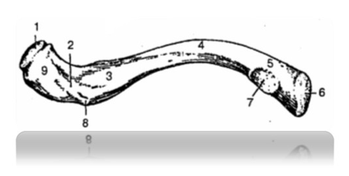
Fig. 81 Right clavicle (bottom view) 528 312 918 222:
1 – acromial articular surface 528 314 818 214
2 – trapezoidal line 428 712 928 222
3 – groove of the subclavian muscle 528 222 918 412
4 – body of the clavicle 314 812 514 812
5 – sternal end 598 712 819 232
6 – sternal articular surface 548 712 918 322
7 – costoclavicular ligament depression 577 489 312 819
8 – cone-shaped tubercle 521 479 811 299
9 – acromial end 578 912 319 228
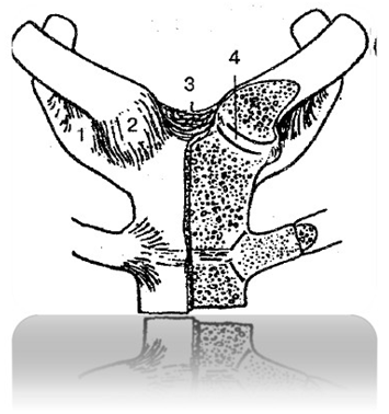
Fig. 82 Sternoclavicular joints 512 714 312 814:
1 – costoclavicular ligament 529 319 819 478
2 – anterior sternoclavicular ligament 319 814 219 217
3 – interclavicular ligament 848 061 219 714
4 – articular disc 519 712 819 912
Skeleton of the free upper limb 219 317 918 817
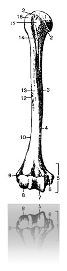
Fig. 83 Right humerus (front view) 524 377 914 877:
1 – head of humerus 219 312 819 778
2 – anatomical neck 219 312 819 887
3 – medial anterior surface 594 716 888 219
4 – medial edge 428 712 328 718
5 – condyle of the humerus 213 428 219 488
6 – medial epicondyle 219 317 819 217
7 – humeral block 539 817 919 817
8 – head of the humeral condyle 219 317 229 812
9 – lateral epicondyle 419 418 712 319
10 – lateral edge 513 814 713 914
11 – body of humerus 548 912 938 817
12 – deltoid tuberosity 548 714 818 217
13 – lateral anterior surface 548 321 918 711
14 – surgical neck 542 718 312 918
15 – small tubercle 328 784 548 914
16 – large tubercle 328 722 588 731
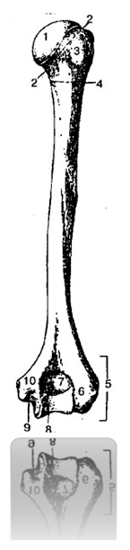
Fig. 84 Right humerus (posterior view)
524 377 914 877:
1 – head of humerus 219 312 819 778
2 – anatomical neck 219 312 819 887
3 – large tubercle 328 722 588 731
4 – surgical neck 542 718 312 918
5 – condyle of the humerus 213 428 219 488
6 – lateral epicondyle 419 418 712 319
7 – elbow joint fossa 519 317 818 907
8 – humeral block 539 817 919 817
9 – groove of the ulnar nerve 298 712 319 212
10 – medial epicondyle 219 317 819 217
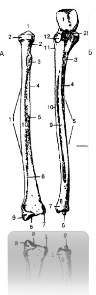
Fig. 85 Right radius and ulna (front view) 598 712 498 217:
A – radius bone 529 321 919 742:
1 – head of the radius 328 471 918 221
2 – neck of the radius 248 312 818 222
3 – tuberosity of the radius 548 712 918 272
4 – interosseous margin 599 614 819 314
5 – front surface 548 371 998 211
6 – front edge 428 317 918 917
7 – elbow notch 498 318 919 887
8 – carpal articular surface 319 817 919 617
9 – styloid process 514 317 914 987
10 – lateral surface 598 712 918 312
11 – body of the radius 598 321 719 811
12 – articular circumference 549 312 919 812
B – ulna 529 341 419 811:
1 - block notch 219 217 919 817
2 – coronoid process 319 374 819 814
3 – ulnar tuberosity 818 906 319 481
4 – front edge 518 614 318 488
5 – body of the ulna 598 417 218 611
6 – styloid process 514 317 914 987
7 – articular circumference 519 342 819 221
8 – head of the ulna 548 711 919 211
9 – front surface 534 217 918 377
10 – interosseous margin 598 321 918 211
11 – instep support comb 498 871 218 321
12 - radial notch 548 388 718 918
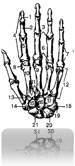
Fig. 86 Bones of the right hand (dorsal surface)
548 722 888 222:
1 – distal phalanx 219 333 819 444
2 – middle phalanx 478 212 978 111
3 – head of the phalanx 513 918 216 097
4 – phalanges (finger bones) 213 918 712 889
5 – proximal phalanx 248 617 319 516
6 – base of phalanx 519 819 312 888
7 – body of the phalanx 891 319 498 519
8 – head of metacarpal bone 519 614 819 310
9 – third metacarpal bone 714 016 218 961
10 – body of metacarpal bone 498 712 988 318
11 – base of metacarpal bone 528 313 818 713
12 – metacarpus (1 – V metacarpal bones) 598 318 488 712
13 – styloid process 528 899 319 217
14 – trapezium bone 988 713 428 755
15 – trapezoid bone 598 317 988 978
16 – capitate bone 298 317 898 007
17 – hamate bone 598 382 488 722
18 – triangular bone 598 213 009 216
19 – pisiform bone 598 377 988 217
20 – lunate bone 598 327 918 227
21 – scaphoid bone 599 891 488 788
Joints of the bones of the upper limb 248 916 319 281
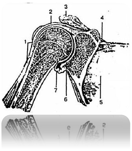
Fig. 87 Shoulder joint (longitudinal view)
219 419 213 818:
1 - tendon of the long head of the biceps muscle
shoulder 529 321 919 478
2, 7 – joint capsule 471 818 321 988
3 – acromion of the scapula 519 312 948 212
4 – superior transverse ligament of the scapula 418 694 398 016
5 – shoulder blade 219 312 988 712
6 – glenoid cavity of the scapula 918 317 988 817
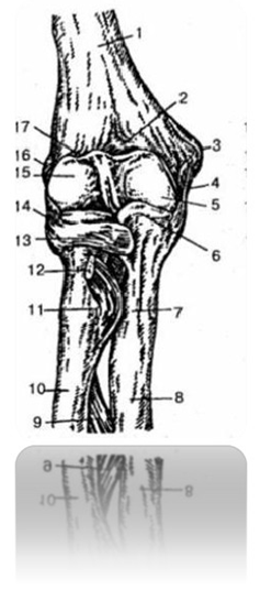
Fig. 88 Right elbow joint (front view)
598 312 419 812:
1 – humerus 524 377 914 877
2 – coronary fossa 599 917 319 878
3 – medial epicondyle 219 317 819 217
4, 13, 14 – bundles 594 316 918 071
5 – humeral block 539 817 919 817
6 – coronoid process 319 374 819 814
7 – ulnar tuberosity 818 906 319 481
8 – ulna 529 341 419 811
9 – interosseous membrane of the forearm 918 312 888 512
10 – radius 529 321 919 742
11 – tuberosity of the radius 548 712 918 272
12 – biceps tendon 528 377 948 724
15 – head of humerus 219 312 819 778
16 – lateral epicondyle 419 418 712 319
17 – radial fossa 529 213 719 333
Bones of the lower limb 529 531 919 811
Lower limb belt 429 712 918 222
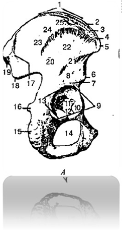
Fig. 89 Pelvic bone, right 214 317 918 227:
A – lateral view:
1 – iliac crest 298 327 918 887
2 – inner lip 291 398 218 612
3 – intermediate line 549 715 819 315
4 – outer lip 599 422 899 322
5 – superior anterior iliac spine 529 312 919 914
6 – inferior anterior iliac spine 598 714 818 914
7 – supraacetabular groove 594 321 714 811
8 – body of the ilium 548 377 914 817
9 – acetabulum edge 519 718 918 212
10 – crescent surface 498 712 519 312
11 – acetabulum fossa 548 712 918 312
12 – acetabulum notch 509 714 219 314
13 – acetabulum 591 614 318 790
14 – locking hole 521 782 219 332
15 – ischial tuberosity 529 312 918 812
16 – ischial spine 898 918 314 517
17 – posterior gluteal line 517 318 215 428
18 – inferior posterior iliac spine 912 914 712 714
19 – superior posterior iliac spine 319 812 892 319
20 – ilium 218 317 228 917
21 – lower gluteal line 519 312 891 421
22 – gluteal surface 519 311 819 211
23 – iliac wing 529 301 229 721
24 – anterior gluteal line 498 312 898 222
25 – iliac tubercle 594 312 894 222

B - view from the medial side:
1 – iliac crest 298 327 918 887
2 – superior posterior iliac spine 319 812 892 319
3 – sacropelvic surface 298 712 518 312
4 – iliac tuberosity 498 213 009 671
5 – inferior posterior iliac spine 912 914 712 714
6 – ear-shaped surface 931 489 219 819
7 – locking hole 521 782 219 332
8 – arc line 512 098 096 681
9 – inferior anterior iliac spine 598 714 818 914
10 – body of the ilium 488 714 818 381
11 – ilium 218 317 228 917
12 – superior anterior iliac spine 529 312 919 914
13 – iliac fossa 489 091 213 719
14 – iliac wing 529 301 229 721
15 – inner lip 291 398 218 612
Skeleton of the free part of the lower limb 298 321 918 557
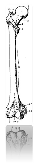
Fig. 90 Right femur (front view)
918 275 784 325:
1 – head of the femur 519 517 919 917
2 – fossa of the head of the femur 319 318 919 818
3 – femoral neck 498 312 718 212
4 – intertrochanteric line 519 312 219 772
5 – small spit 918 714 518 914
6 – body of the femur 489 061 398 719
7 – adductor tubercle 498 312 000 612
8 – medial epicondyle 469 718 919 318
9 – medial condyle 598 317 898 617
10 – patellar surface 514 318 914 888
11 – lateral condyle 598 314 888 914
12 – lateral epicondyle 519 714 814 919
13 – Large spit 529 318 729 888
14 – trochanteric fossa 598 712 888 412
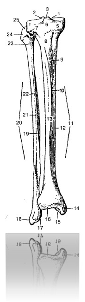
Fig. 91 Right tibia and fibula (anterior view)
519 714 479 509:
1 – lateral condyle 519 514 319 314
2 – lateral intercondylar tubercle 539 712 819 222
3 – intercondylar eminence 548 917 328 227
4 – medial intercondylar tubercle 219 713 829 223
5 – medial condyle 394 815 519 815
6 – anterior intercondylar field 591 488 219 181
7 – articular surface of the head of the fibula
598 617 298 319
8 – tibial tuberosity 529 327 819 227
9 – medial surface 548 912 914 272
10 – front edge 428 319 819 228
11 – body of the tibia 829 714 329 214
12 – medial edge 314 812 914 712
13 – lateral surface 519 312 914 212
14 – medial malleolus 589 741 299 421
15 – articular surface of the ankle 528 714 328 214
16 – lower articular surface 594 321 317 811
17 – articular surface of the malleolus of the fibula 598 712 918 241
18 – lateral malleolus of the fibula 514 718 914 318
19 – medial surface of the fibula 489 916 769 817
20 – body of fibula 538 714 918 214
21 – anterior edge of fibula 298 718 314 228
22 – lateral surface of the fibula
298 317 918 227
23 – neck of fibula 238 714 214 816
24 – head of fibula 519 317 919 221
25 – apex of the head of the fibula 518 318 918 227
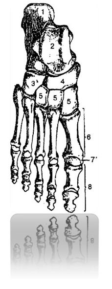
Fig. 92 Bones of the right foot (top view) 594 317 214 817:
1 – calcaneus 594 312 814 712
2 – talus 594 317 814 227
3 – cuboid bone 598 317 998 217
4 – scaphoid bone 598 317 918 228
5 – sphenoid bones 284 318 914 278
6 – metatarsal (I – V) bones 918 714 888 914
7 – head of metatarsal bone 538 717 918 227
8 – finger bones (proximal, middle and distal)
phalanges) 598 317 888 999
Joints of the bones of the lower limb 918 999 000 818
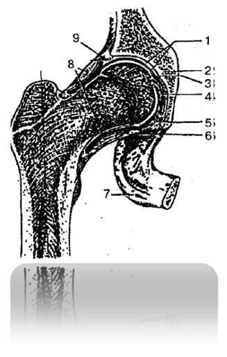
Fig. 93 Hip joint (right) 319 489 219 318:
1 – articular cartilage 219 914 319 814
2 – pelvic bone 598 019 319 181
3 – joint cavity 519 318 919 888
4 – ligament of the head of the femur 548 317 228 917
5 – transverse acetabular ligament 514 318 814 228
6 – joint capsule 987 421 328 921
7 – ischial tuberosity 529 312 918 812
8 – circular zone 598 712 998 212
9 – acetabular labrum 219 317 559 417
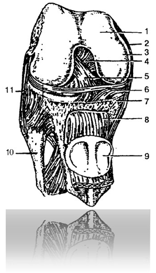
Fig. 94 Knee joint 419 718 214 328:
1 – femur 918 275 784 325
2 – tibial collateral ligament 548 617 019 311
3 – medial condyle 498 712 919 812
4 – posterior cruciate ligament 419 321 819 221
5 – anterior cruciate ligament 219 314 919 814
6 – medial meniscus 928 601 598 219
7 – tibia 918 321 989 711
8 – patellar ligament 594 312 894 882
9 – patella 421 891 529 328
10 – fibula 498 217 888 917
11 – fibular collateral ligament 529 317 889 227
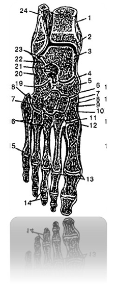
Fig. 95 Joints and ligaments of the right foot 419 417 819 817:
1 – tibia 918 321 989 711
2 – ankle joint cavity 429 327 919 877
3, 7, 12, 13, 16, 18, 19, 20, 21, 23 – ligaments that strengthen
joints 510 648 219 310
4 – transverse joint of the tarsus 391 819 291 919
5 – scaphoid bone 598 317 918 228
6 – cuneonavicular joint 514 387 914 327
8, 9, 10 – sphenoid bones 284 318 914 278
11 – tarsometatarsal joints 519 518 919 818
14 – interphalangeal joints 518 716 319 498
15 – metatarsophalangeal joint (V) 594 312 814 212
17 – cuboid bone 598 317 998 217
22 – subtalar joint 594 317 814 217
24 – fibula 498 217 888 917
MUSCULAR SYSTEM 214 712 314 222
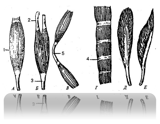
Fig. 96 Numerical concentrations by form
muscles 898 811 919 218:
A - fusiform muscle 319 241 809 217
B – biceps 914 312 219 312
B - digastric muscle 214 318 914 718
G - muscle with tendinous bridges
519 317 914 817
D – bipennate muscle 498 712 319 212
E - unipennate muscle 519 314 219 814
1 – belly muscle 719 618 914 217
2, 3 – tendons of muscle 598 317 918 227
4 – tendon bridge 519 641 319 071
5 – intermediate tendon 319 919 819 318
Concentrations on numbers that restore the form of matter allow one to apply the corresponding
concentrations of the spiritual state for the restoration of the shape of individual organs and the entire organism.
Muscles and fascia of the trunk 519 614 819 317
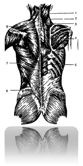
Fig. 97 Superficial muscles of the back 819 314 914 812:
1 – splenius capitis muscle 298 742 279 488
2 – muscle that lifts the scapula 214 317 914 717
3 – rhomboid minor muscle 319 061 919 618
4 – large rhomboid muscle 584 317 914 016
5 – lower posterior serratus muscle 549 317 919 817
6 – lumbar-fugal fascia 529 317 919 817
7 – latissimus dorsi 429 318 829 998
8 – trapezius muscle 421 317 921 817
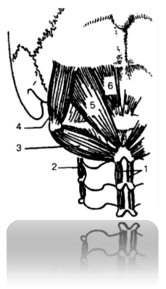
Fig. 98 Suboccipital muscles 531 814 212 814:
1 – interspinous muscles of the neck 489 617 819 398
2 – intertransverse muscles 519 314 819 312
3 – inferior oblique muscle of the head 218 317 918 227
4 – superior oblique muscle of the head 218 417 918 817
5 – large posterior rectus capitis muscle 594 318 614 715
6 – rectus capitis posterior minor 219 817 819 227
Back fascia 214 718 314 888
1 – pectoralis major 214 718 918 228
2 – pectoralis minor muscle (muscle contours) 521 618 919 192
3 – anterior serratus muscle 219 475 819 355
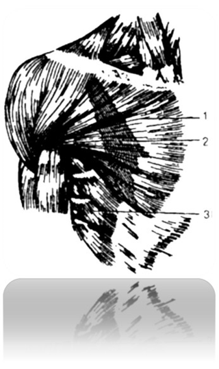
Fig. 99 Chest muscles 489 611 098 217:
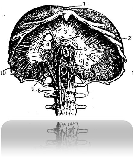
Fig. 100 Diaphragm (bottom view) 219 289 228 317:
1 – sternal part of the diaphragm 519 517 819 217
2, 10 – costal part of the diaphragm 214 312 489 212
3 – tendon center 528 974 319 641
4 – opening for the inferior vena cava 519 518 919 218
5 – opening for the esophagus 528 317 918 227
6 – aortic opening 548 312 918 227
7, 8, 9 – legs of the lumbar part of the diaphragm
549 316 860 219
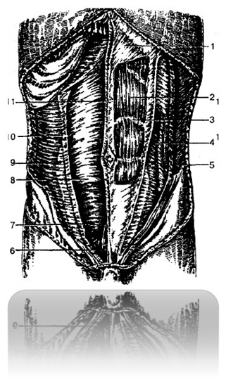
Fig. 101 Abdominal muscles 517 318 917 918:
1 – aponeurosis of the external oblique muscle of the abdomen 498 641 819 071
2 – rectus abdominis 598 712 319 217
3 – tendon bridge 528 714 328 917
4 – internal oblique muscle of the abdomen 398 217 818 417
5 – external oblique abdominal muscle 529 312 419 272
6 – pyramidal muscle 598 742 218 228
7, 8 – posterior wall of the rectus abdominis sheath
213 019 514 219
9 – aponeurosis of the transverse abdominal muscle 418 714 918 444
10 – transverse abdominal muscle 555 813 915 513
11 – white line of the abdomen 319 481 219 321
Muscles and fascia of the head and neck 219 214 419 314
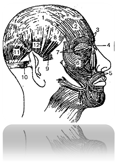
Fig. 102 Superficial facial muscles
heads 219 317 914 817:
1 – tendon helmet 314 812 914 212
2 – occipitofrontalis muscle 009 812 214 312
3, 6, 7 – orbicularis oculi muscle 214 317 914 217
4 – muscle of the proud 829 312 714 712
5 – orbicularis oris muscle 548 321 818 221
8 – temporoparietal muscle 409 641 219 741
9 – anterior auricular muscle 519 312 518 222
10 – posterior auricular muscle 548 314 214 317
11 – occipitofrontalis muscle 009 812 214 312
12 – superior auricularis muscle 519 812 919 912
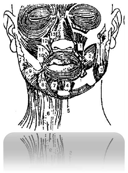
Fig. 103 Facial muscles (front view)
598 641 398 719:
1 - muscle that raises the angle of the mouth 598 712 918 242
2 – cheek muscle 549 317 849 217
3 – chewing muscle 598 712 918 212
4 – mentalis muscle 314 312 814 212
5 – transverse chin muscle 519 784 398 419
6 – subcutaneous muscle of the neck 518 317 918 227
7 – muscle that depresses the lower lip 521 319 221 919
8 – muscle that depresses the angle of the mouth 518 519 318 919
9 – laughter muscle 418 716 818 999
10 – zygomaticus major muscle 548 712 998 218
11 – zygomaticus minor muscle 598 217 918 227
12 – muscle that raises the upper lip 319 817 219 227
13 – muscle that lifts the upper lip and wing of the nose
419 227 299 327
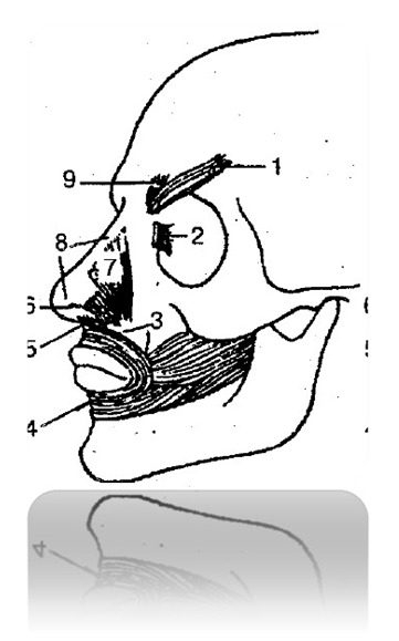
Fig. 104 Deep facial muscles 328 721 428 919:
1 – corrugator supercilii muscle 512 428 912 728
2 – lacrimal part of the orbicularis oculi muscle 319 742 819 222
3 – marginal part of the orbicularis oris muscle 548 232 619 722
4 – labial part of the orbicularis oris muscle 428 713 918 523
5 – muscle that depresses the septum of the nose 521 216 719 226
6 – alar part of the nasal muscle 428 921 321 481
7 – transverse part of the nasal muscle 219 317 819 228
8 – nasal muscle 498 712 518 232
9 – muscle that lowers the eyebrow 514 217 914 217
Masticatory muscles 519 314 819 214
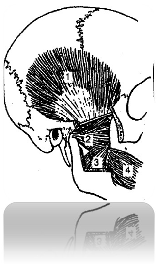
Fig. 105 Masticatory muscles 519 314 819 214:
1 – temporal muscle 218 317 918 217
2 – lateral pterygoid muscle 219 214 319 214
3 – medial pterygoid muscle 819 912 314 272
4 – cheek muscle 549 317 849 217
Fascia of the head 519 718 218 314
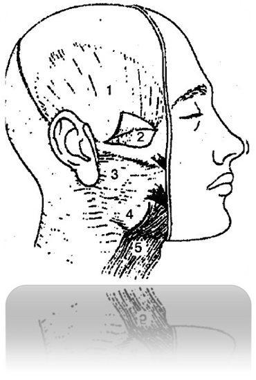
Fig. 106 Fasciae of the head 519 718 218 314:
1 – temporal fascia 319 814 919 214
2 – deep plate of the temporal fascia 498 641 319 712
3 – fascia of the parotid gland 519 814 719 314
4 – chewing fascia 898 716 214 319
5 – subcutaneous muscle of the neck 518 317 918 227

Neck muscles 548 007 998 227
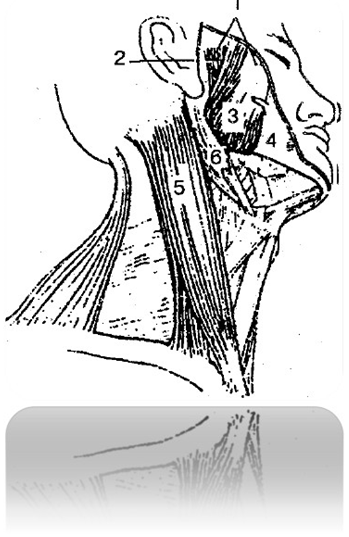
Fig. 107 Muscles of the head and neck (right and bottom view)
528 342 918 712:
1 – chewing muscle 598 712 918 212
2 – deep part of the masseter muscle 574 321 914 211
3 – superficial part of the masseter muscle 584 312 984 212
4 – buccal-pharyngeal fascia 217 317 519 715
5 – sternocleidomastoid muscle 548 712 218 312
6 – digastric muscle 214 318 914 718

Deep Neck Muscles 819 341 919 841
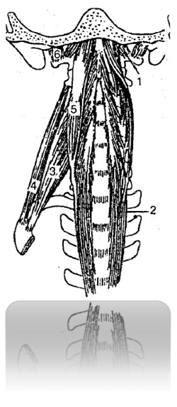
Fig. 108 Deep muscles of the neck 819 341 919 841:
1 – anterior rectus capitis muscle 519 642 068 917
2 – long muscle of the neck 213 418 913 818
3 – anterior scalene muscle 599 681 917 298
4 – middle scalene muscle 489 316 718 916
5 – longus capitis 519 817 319 817
6 – lateral rectus capitis 498 061 319 789
Muscles and fascia of the upper limb 219 314 819 914
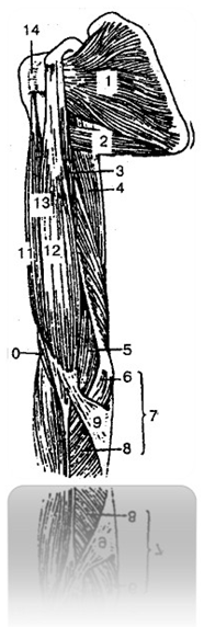
Fig. 109 Muscles of the girdle of the upper limb and shoulder (front view)
498 711 598 321:
1 – subscapularis muscle 519 312 219 282
2 – large round muscle 459 321 989 721
3 – coracobrachialis muscle 219 371 419 871
4 – triceps brachii 419 812 599 322
5, 10 – shoulder muscle 319 717 819 317
6 – humeral head of the round pronator 598 317 819 217
7 – round pronator 214 888 219 317
8 – ulnar head of the round pronator 919 814 319 214
9 – aponeurosis of the biceps brachii muscle 428 312 818 512
11 – long head of the biceps brachii 519 817 919 017
12 – short head of the biceps brachii 584 712 584 389
13 – biceps brachii 549 817 319 217
14 – intertubercular sheath of the tendon of the long head of the biceps muscle 214 987 914 317
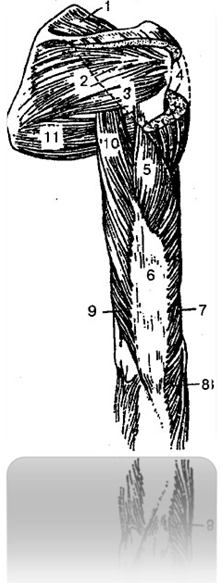
Fig. 110 Muscles of the girdle of the upper limb and shoulder (posterior view)
498 711 598 321:
1 – supraspinatus muscle 312 214 812 514
2 – infraspinatus muscle 548 619 319 071
3 – small round muscle 555 333 918 433
4 – deltoid muscle 598 371 888 911
5 – lateral head of the triceps brachii 598 909 913 718
6 – triceps brachii 419 812 599 322
7, 9 – medial head of the triceps brachii
529 382 728 814
8 – elbow muscle 517 214 917 814
10 – long head of the triceps brachii 512 718 912 229
11 – large round muscle 459 321 989 721

Fig. 111 Forearm muscles (anterior group) 798 064 219 317:
1 – aponeurosis of the biceps brachii muscle 428 312 818 512
2 – biceps brachii 549 817 319 217
3 – round pronator 214 888 219 317
4 – long palmar muscle 498 715 318 225
5 – Flexor carpi ulnaris 498 094 319 591
6 – humero-ulnar head of the superficial flexor of the fingers 513 819 913 514
7 – superficial flexor of the fingers 319 811 919 891
8 – short palmar muscle 598 319 719 818
9 - Square Pronator 489 016 718 941
10 – radial head of the superficial flexor of the fingers
598 317 588 817
11 – long radial extensor of the wrist 598 214 319 814
12 – flexor carpi radialis 511 408 219 319
13 – brachioradialis muscle 429 318 229 718

Fig. 112 Muscles of the right hand (front view) 214 717 814 327:
1 – flexor retainer 519 321 019 614
2 – muscle abducting the little finger 318 912 818 006
3 – short flexor of the little finger 519 388 498 514
4 – tendons of the deep flexor of the fingers 588 317 918 777
5 – opposing muscle of the little finger 988 888 314 007
6 – lumbrical muscles 568 721 328 521
7 – tendons of the superficial flexor of the fingers 598 712 899 422
8 – muscle that adducts the thumb of the hand 599 712 899 329
9 – tendon of the long flexor of the thumb
548 916 379 816
10 – short flexor of the pollicis 898 317 879 917
11 – short muscle that abducts the thumb of the hand
389 671 899 211
Muscles of the lower limb 514 311 914 527
Pelvic muscles 298 317 919 817

Fig. 113 Muscles of the lower limb 514 311 914 527:
A - front view: 1 - iliopsoas muscle 319 311 919 811
2 – pectineus muscle 519 712 918 412
3 – long adductor muscle 519 312 819 712
4 – thin muscle 319 042 219 822
5 – sartorius muscle 899 389 919 388
6 – medial vastus muscle 988 091 891 491
7 – quadriceps tendon 514 517 814 317
8 – patellar ligament 594 312 894 882
9 – gastrocnemius muscle 589 319 814 228
10 – soleus muscle 498 513 818 933
11 – long extensor of the fingers 318 512 818 064
12 – long peroneus muscle 528 426 918 726
13 – anterior tibialis muscle 548 717 318 917
14 – lateral vastus muscle 548 964 328 744
15 – rectus femoris 589 371 841 218
B – back view: 1 – gluteus maximus 498 614 319 214
2 – iliotibial tract (part of the broad fascia of the thigh)
219 371 819 511
3 – biceps femoris 598 617 329 817
4 – gastrocnemius muscle 589 319 814 228
5 – Achilles tendon 598 317 418 917
6 – semimembranosus muscle 388 427 918 227
7 – semitendinosus muscle 549 381 714 817
Muscles of the free part of the lower limb 319 715 819 555
Calf muscles 329 481 918 511
Muscles of the foot 519 371 819 511
Fascia of the lower limb 529 377 429 879
Fascia of the leg 539 427 819 677
INTERNAL ORGANS 523,000,898,111
Digestive system 541 928 741 588
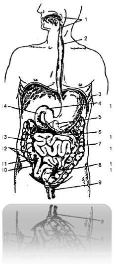
Fig. 114 Digestive tract 514 388 914 888:
1 - pharynx 519 987 319 427
2 – esophagus 598 381 698 711
3 – stomach 898 898 478 213
4 – the transition point of the stomach into the duodenum
598 513 998 719
5 - the transition point of the duodenum into the jejunum
214 511 819 311
6 – jejunum 548 714 318 215
7 – descending colon 248 389 428 999
8 – sigmoid colon 319 812 519 427
9 – rectum 598 714 898 314
10 – vermiform appendix 529 317 899 228
11 – ileum 394 897 594 377
12 – cecum 519 489 319 788
13 – ascending colon 599 213 988 713
14 – duodenum 589 608 488 914
Oral cavity 891 000 499 887
Oral glands 319 841 519 811
Throat 519 987 319 427
Esophagus 598 381 698 711
Stomach 898 898 478 213
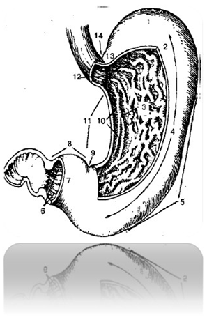
Fig. 115 Stomach 898 898 478 213:
1 – fundus of the stomach 898 319 899 214
2 – front wall 514 878 917 887
3 – folds of the stomach 598 317 918 527
4 – body of the stomach 889 919 389 418
5 – greater curvature of the stomach 489 981 948 513
6 – Gatekeeper channel 518 917 319 877
7 – Gatekeeper's Cave 548 711 919 411
8 – pyloric part 591 488 791 888
9 - Corner Cut 596 317 549 817
10 – stomach channel 599 481 799 811
11 – lesser curvature of the stomach 418 728 319 348
12 – cardiac orifice 528 481 798 711
13 – cardiac part of the stomach 521 316 891 714
14 – cardiac notch 517 916 815 322
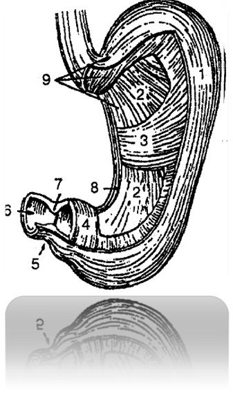
Fig. 116 Muscular membrane of the stomach 491 516 018 329:
1, 8 – longitudinal layer 599 891 799 811
2 – oblique fibers 534 981 814 917
3, 4 – circular layer 599 317 819 417
5 – gatekeeper 598 421 918 511
6 – gatekeeper hole 598 714 818 314
7 – pyloric sphincter 598 714 918 324
9 – muscular membrane 584 321 844 711
Small intestine 528 317 428 717
Liver, duodenum, pancreas 219 214 319 714
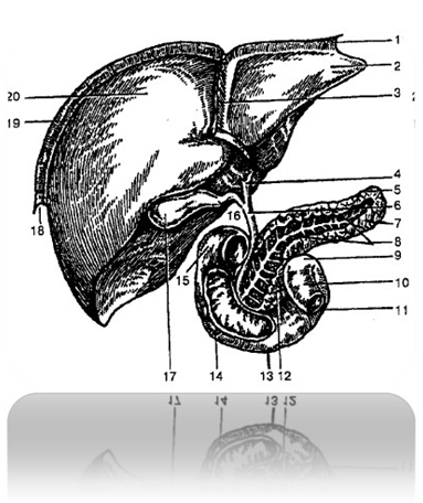
Fig. 117 Liver, duodenum, pancreas
iron 219 214 319 714:
1 – left triangular ligament 519 617 919 817
2 – left lobe of the liver 988 411 218 217
3 – falciform ligament (of the liver) 214 311 714 811
4 – common hepatic duct 914 311 814 019
5 – pancreas 589 317 919 877
6 – common bile duct 599 381 989 391
7 – tail of pancreas 898 429 719 482
8 – pancreatic duct 599 316 739 928
9 – duodenojejunal flexure 214 279 881 319
10 – jejunum 548 714 318 215
11 – ascending duodenum 591 477 391 817
12 – head of pancreas 319 487 914 917
13 – horizontal part of the duodenum
519 314 819 414
14 – descending part of the duodenum 019 819 319 417
15 – upper part of the duodenum 519 811 919 311
16 – cystic duct 599 811 919 891
17 – Gallbladder 918 712 418 912
18 – right triangular ligament 588 918 319 819
19 – coronary ligament 514 312 914 812
20 – right lobe of the liver 598 614 298 719
Large intestine 591 488 898 217
Abdominal cavity and peritoneum 598 123 098 719
RESPIRATORY SYSTEM 598 788 428 317
Nasal cavity 428 317 818 227
Larynx 291 891 419 391
Laryngeal cartilages 529 319 489 518
Muscles of the larynx 594 318 719 214
Laryngeal cavity 581 398 421 898

Fig. 118 Laryngeal cavity (frontal view) 581 398 421 898:
1 – epiglottis 214 317 814 817
2 – epiglottic tubercle 219 329 814 718
3 – vestibule of the larynx 519 481 299 811
4 – vestibule fold 599 016 719 317
5 – laryngeal ventricle 528 391 919 811
6 – vocal fold 598 718 319 421
7 – thyroid cartilage 588 421 388 711
8 – glottis 528 742 318 014
9 – subglottic cavity 514 781 910 094
10 – tracheal cavity 298 719 488 919
11 – cricoid cartilage 584 317 589 307
12 – lateral cricoarytenoid muscle 219 387 919 227
13 – vocal muscle 594 817 914 919
14 – thyroarytenoid muscle 513 819 014 912
15 – vestibule slit 598 741 998 328
Trachea and bronchi 428 714 008 914
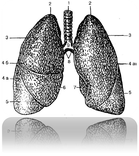
Fig. 119 Trachea, main bronchi and lungs
891 321 511 981:
1 – trachea 429 318 919 888
2 – apex of the lung 598 061 319 212
3 – upper lobe 529 312 547 399
4 a – oblique gap 498 712 818 918
4 b – horizontal gap 819 321 918 898
5 – lower lobe 214 318 718 912
6 – average share 519,812,919,422
7 – cardiac notch of the left lung 528 316 398 641
8 – main bronchi 819 314 899 049
9 – bifurcation of the trachea 514 518 314 818
Lungs 519 418 319 818
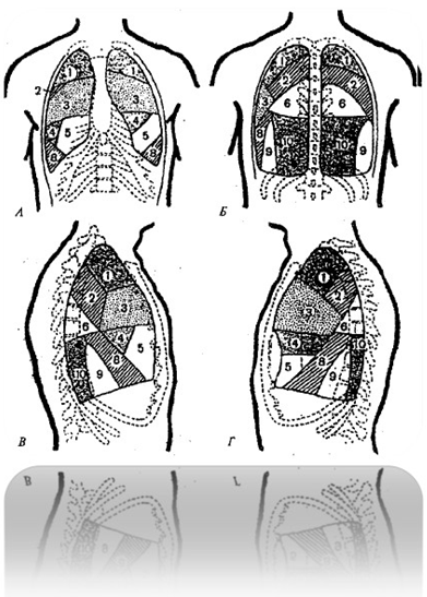
Fig. 120 Segments of the lung:
A - front view
B - rear view
B - right lung (lateral view)
G – left lung (lateral view)
1 segment 914 818 312 898
2 segment 319 814 919 914
3 segment 219 318 919 818
4 segment 519 319 818 214
5 segment 918 319 819 212
6 segment 314 819 888 915
7 segment 219 319 812 794
8 segment 319 419 898 912
9 segment 319,892 219,844
10 segment 319 488 988 210
Pleura and mediastinum 898 315 428 188
UROGENITAL APPARATUS 898 398 412 842
Kidney 289 391 814 216
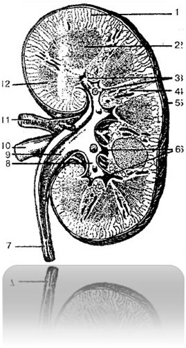
Fig. 121 Right kidney (frontal view)
289 391 814 216:
1 – cortex 912 898 949 319
2 – brain substance 598 716 399 008
3 – renal papillae 519 377 918 227
4 – renal columns 594 312 898 714
5 – fibrous capsule 980 841 218 329
6 – minor renal calyces 589 326 799 816
7 – ureter 214 312 810 008
8 – large renal calyx 989 217 519 487
9 – renal pelvis 928 899 499 711
10 – renal vein 518 914 319 528
11 – renal artery 198 711 298 241
12 – renal pyramid 539 871 491 001
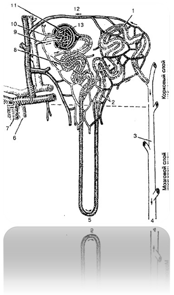
Fig. 122 Structure and blood supply of the nephron:
1 – distal convoluted tubule 814 988 719 081
2 – capillary network 214 718 918 317
3 – collecting duct 548 219 317 917
4 – movement of urine to the renal pelvis 298 017 498 248
5 – loop of Henle 518 319 888 910
6 – renal artery 198 711 298 241
7 – renal vein 518 914 319 528
8 – proximal convoluted tubule 571 898 714 557
9 – afferent arteriole 889 014 219 893
10 – efferent arteriole 890 014 318 714
11 – renal glomerulus 519 891 249 318
12 – venule 213 984 791 248
13 – Bowman’s capsule 298 788 489 791
Ureters 214 312 810 008
Bladder 219 389 998 419
Urethra 329 487 948 216
Male genitalia 519 007 898 367
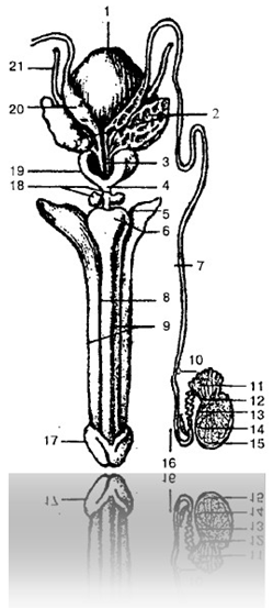
Fig. 123 Internal and external male genitalia:
1 – urinary bladder 219 389 998 419
2 – seminal vesicle 519 317 898 487
3 – ejaculatory duct 591 488 011 228
4 – membranous part of the urethra 319 487 919 008
5 – leg of the penis 819 317 919 847
6 – bulb of the penis 528 719 048 317
7 – vas deferens 398 755 819 455
8 – corpus spongiosum 398 787 914 321
9 – corpus cavernosum 598 721 598 311
10 – epididymis 519 488 299 318
11 – efferent tubules 548 371 818 211
12 – testicular mesh 938 729 488 219
13 – straight seminiferous tubules 419 871 989 311
14 – convoluted seminiferous tubules 498 712 819 212
15 – protein shell 319 817 919 217
16 – lower part of the vas deferens 519 891 499 217
17 – head of the penis 528 714 314 712
18 – bulbourethral gland 519 712 419 812
19 – prostate gland 498 714 918 214
20 – ampulla of the vas deferens 418 918 971 998
21 – ureter 214 312 810 008
Female genitals 519 814 089 319
Internal female genital organs 419 219 808 319
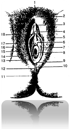
Fig. 124 Female external genitalia 519 319 818 678:
1 – pubis 519 317 898 498
2 – anterior commissure of the lips 591 489 719 328
3 – foreskin of the clitoris 419 319 898 987
4 – head of the clitoris 980 409 501 201
5 – labia majora 598 711 008 512
6 – paraurethral ducts 598 641 788 910
7 – labia minora 319 016 789 498
8 – duct of the large gland of the vestibule 889 014 317 489
9 – frenulum of the labia 598 021 318 714
10 – posterior commissure of the lips 539 421 819 317
11 – anus 589 317 418 917
12 – perineum 398 711 419 411
13 – fossa of the vestibule of the vagina 589 471 219 889
14 – hymen 529 314 789 064
15 – vaginal opening 591 472 918 223
16 – vestibule of the vagina 888 017 989 117
17 – external opening of the urethra
498 663 219 773
18 – frenulum of the clitoris 398 421 891 871
ENDOCRINE GLANDS 889 314 219 798
Pituitary and pineal gland 214 318 908 210
Pituitary gland 317 218 219 819
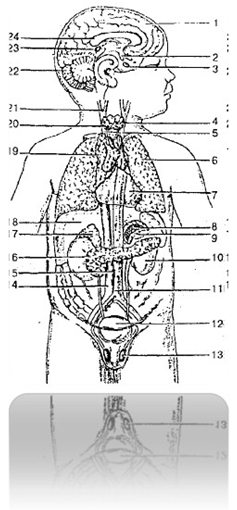
Fig. 125 Location of human endocrine glands:
1 – hemisphere of the cerebrum 819 917 819 319
2 – hypothalamic nuclei 890 498 319 718
3 – pituitary gland 317 218 219 819
4 – thyroid gland 829 319 409 819
5 – trachea 429 318 919 888
6 – light 519 418 319 818
7 – pericardium 989 387 988 878
8 – adrenal medulla 549 378 918 268
9 – adrenal cortex 912 898 949 319
10 – kidney 289 391 814 216
11 – aorta 398 071 890 498
12 – urinary bladder 219 389 998 419
13 – testicle 298 017 319 487
14 – inferior vena cava 549 671 919 871
15 – aortic paraganglion 591 488 018 713
16 – pancreas 589 317 919 877
17 – adrenal gland 891 418 712 319
18 – liver 219 712 919 222
19 – thymus gland 481 914 319 814
20 – parathyroid gland 219 319 895 219
21 – Sleepy Glomus 549 641 898 017
22 – cerebellum 828 219 328 299
23 – pineal gland (epiphysis) 489 641 399 042
24 – corpus callosum 498 712 328 071
Numerical concentrations on endocrine glands by their location areas can be produced by successively concentrating on the numbers corresponding to endocrine glands 889 314 219 798 and on the numbers of organs in the area of which endocrine glands are located. This method can be used in the principle of restoration by numerical concentrations, in which, along with the restoration of the selected tissue, it is advisable to restore the areas adjacent to the tissue.
Numerical series of regulation of endocrine glands
519 317 219 416 can be used to speed up
restoration of matter.
Pineal body (Epiphysis) 489 641 399 042
Thyroid and parathyroid glands 219 318 219 471
Thymus gland 481 914 319 814
Thyroid gland 829 319 409 819
Parathyroid glands 219 319 895 219
Adrenal gland 891 418 712 319
Endocrine pancreas 918 712 818 229
Endocrine part of the sex glands 519 318 914 019
Regulation of endocrine glands 519 317 219 416
CARDIOVASCULAR SYSTEM 214 700 819 891
Arteries, veins and capillaries 219 387 919 887
Heart 918 749 328 081
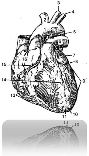
Fig. 126 Heart (front view) 918 749 328 081:
1 – aorta 398 071 890 498
2 – brachiocephalic trunk 998 301 248 227
3 – left common carotid artery 428 712 488 913
4 – left subclavian artery 429 387 219 377
5 – arterial ligament 519 481 319 818
6 – pulmonary trunk 519 421 819 221
7 – left ear 519 318 219 481
8, 15 – coronal groove 519 312 814 829
9 – left ventricle 589 348 914 918
10 – apex of the heart 519 421 899 321
11 – notch of the apex of the heart 528 944 988 714
12 – sternocostal (anterior) surface of the heart
519 317 988 547
13 – right ventricle 598 371 988 011
14 – anterior interventricular groove 909 817 398 787
16 – right ear 598 714 321 898
17 – superior vena cava 398 712 988 012
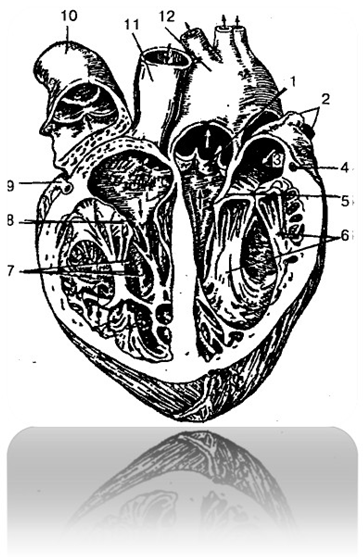
Fig. 127 Heart (open) 918 749 328 081:
1 – semilunar flaps of the aortic valve 591 487 919 877
2 – pulmonary veins 309 428 519 421
3 – left atrium 518 712 314 887
4, 9 – coronary arteries 519 817 319 487
5 – left atrioventricular (mitral) valve
(Bi-leaf valve) 598 517 818 617
6 – papillary muscles 598 717 918 317
7 – right ventricle 598 371 988 011
8 – right atrioventricular (tricuspid)
valve 389 412 819 322
10 – pulmonary trunk 519 421 819 221
11 – superior vena cava 398 712 988 012
12 – aorta 398 071 890 498
Conduction system of the heart 989 808 884 318
Blood supply and innervation of the heart 891 318 910 488
Vessels of the pulmonary circulation 898 714 988 569
Pulmonary veins 309 428 519 421
Vessels of the systemic circulation 598 716 588 317
Branches of the aortic arch 298 714 319 814

Fig. 128 Arteries of the head and neck (right view)
518 422 819 312:
1 – dorsal nasal artery 398 718 989 061
2 – infraorbital artery 898 048 319 061
3 – angular artery 288 919 069 789
4 – superior labial artery 598 712 819 328
5 – inferior labial artery 219 318 488 519
6 – submental artery 219 319 489 555
7 – facial artery 219 061 234 890
8 – lingual artery 498 519 401 209
9 – superior thyroid artery 519 513 719 313
10 – common carotid artery 894 317 212 847
11 – inferior thyroid artery 518 377 918 478
12 – superficial artery of the neck 214 381 918 918
13 – thyrocervical trunk 519 317 919 288
14 – subclavian artery 594 712 819 017
15 – suprascapular artery 529 317 419 817
16 – transverse artery of the neck 519 894 512 319
17 – internal carotid artery 549 712 810 248
18 – superficial temporal artery 519 016 319 417
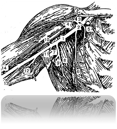
Fig. 129 Arteries of the right axillary fossa
and shoulder 529 317 919 227:
1 – axillary artery 539 891 319 988
2 – thoracoacromial artery 981 516 719 312
3 – acromial branch 214 328 712 918
4 – deltoid branch 594 716 018 988
5 – thoracic branches 317 498 614 219
6 – lateral thoracic artery 598 722 918 213
7 – subscapular artery 598 718 419 087
8 – thoraco-dorsal artery 594 715 319 812
9 – circumflex scapular artery 897 314 421 899
10 – anterior circumflex humeral artery
599 816 719 817
11 – posterior circumflex humeral artery
219 714 819 814
12 – deep brachial artery 317 818 917 918
13 – superior ulnar collateral artery
891 047 089 517
14 – brachial artery 598 641 389 071
Branches of the thoracic aorta 548 617 908 614
Branches of the abdominal aorta 548 711 488 211
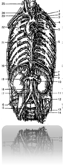
Fig. 130 Thoracic and abdominal aorta 898 121 319 711:
1 – left common carotid artery 428 712 488 913
2 – left subclavian artery 429 387 219 377
3 – internal thoracic artery 598 341 818 941
4 – aortic arch 219 877 549 277
5 – bronchial branches 619 321 819 221
6 – descending aorta 519 817 218 217
7 – celiac trunk 594 315 894 715
8 – superior mesenteric artery 398 712 888 422
9 – diaphragm 219 289 228 317
10 – abdominal aorta 598 316 488 916
11 – inferior mesenteric artery 598 361 988 712
12 – common iliac artery 898 531 314 717
13 – external iliac artery 819 415 919 215
14 – internal iliac artery 584 319 914 899
15 – median sacral artery 598 713 818 213
16 – iliolumbar artery 519 388 918 916
17 – lumbar artery 489 712 319 272
18 – ovarian artery 519 648 319 788
19 – right renal artery 198 711 298 241
20 – inferior phrenic artery 598 318 918 999
21 – intercostal artery 548 316 689 766
22 – ascending aorta 598 712 898 612
23 – brachiocephalic trunk 998 301 248 227
24 – right subclavian artery 598 317 819 227
25 – right common carotid artery 919 421 818 728

Fig. 131 Arteries of the leg 319 421 919 724:
A - front view:
1 – knee joint network 518 471 219 140
2 – tendon of the anterior tibialis muscle
519 717 919 817
3 – tendon of the long extensor of the fingers 319 488 519 318
4 – dorsal artery of the foot 481 319 409 816
5 – long extensor of the thumb 529 361 819 711
6 – long peroneus muscle 528 426 918 726
7 – long extensor of the fingers 318 512 818 064
8 – anterior tibial artery 219 488 714 918
9 – knee joint bag 589 412 919 812
B – rear view:
1 – popliteal artery 988 612 818 719
2 – lateral superior genicular artery 548 321 748 244
3, 10 – gastrocnemius arteries 549 365 814 775
4 – lateral inferior genicular artery 528 312 718 422
5 – posterior tibial recurrent artery 598 711 989 321
6 – anterior tibial artery 219 488 714 918
7 – peroneal artery 594 782 914 882
8 – posterior tibial artery 589 766 914 861
9 – medial inferior genicular artery 595 814 315 914
11 – medial superior genicular artery 514 317 814 919
Veins of the systemic circulation 419 641 819 317
Veins of the heart 891 428 319 298
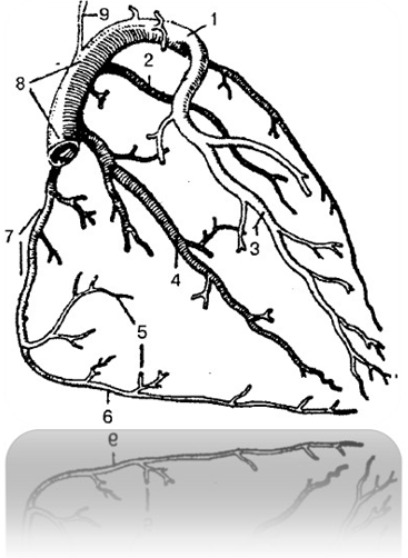
Fig. 132 Veins of the heart 891 428 319 298:
1 – left coronary vein 514 816 718 316
2 – posterior vein of the left ventricle 429 318 719 888
3 – anterior interventricular vein 548 712 918 232
4 – posterior interventricular vein 548 716 328 916
5 – anterior vein of the right ventricle 548 213 898 263
6 – right marginal vein 597 361 326 891
7 – small cardiac vein 598 712 918 322
8 – coronary sinus 578 916 219 316
9 – oblique vein of the left atrium 598 714 319 814
Superior vena cava system 898 317 419 217
Veins of the head and neck 598 716 319 816
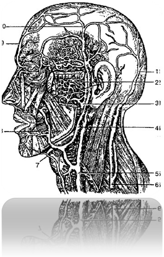
Fig. 133 Veins of the head and face 519 317 919 217:
1 – occipital vein 914 712 298 267
2 – pterygoid (venous) plexus 591 248 791 260
3 – maxillary vein 598 314 818 914
4 – submandibular vein 898 314 718 914
5 – internal jugular vein 598 612 719 322
6 – external jugular vein 594 716 814 516
7 – submental vein 598 714 318 914
8 – facial vein 599 715 819 316
9 – frontal vein 598 781 428 677
10 – superficial temporal vein 548 327 918 227
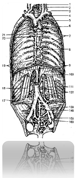
Fig. 134 Veins of the thoracic and abdominal cavities
514 715 914 315:
1 – internal jugular vein 598 612 719 322
2 – external jugular vein 594 716 814 516
3 – subclavian vein 519 371 919 871
4 – brachiocephalic vein 219 378 919 278
5 – superior vena cava 398 712 988 012
6 – bronchi 519 318 619 228
7 – intercostal veins 549 716 919 226
8 – hemiazygos vein 529 317 818 227
9 – diaphragm 219 289 228 317
10 – beginning of the hemiazygos vein 548 712 898 326
11 – inferior vena cava 549 671 919 871
12 – lumbar veins 589 712 919 261
13 – common iliac vein 548 713 918 781
14 – median sacral vein 598 717 318 917
15 – internal iliac vein 549 316 814 787
16 – external iliac vein 999 888 719 898
17 – iliolumbar vein 548 791 018 216
18 – square lumbar muscle 019 321 068 911
19 – beginning of the azygos vein 519 217 918 757
20 – azygos vein 729 329 898 888
21 – accessory hemiazygos vein 818 888 068 712
Inferior vena cava system 394 617 014 809
Portal vein system 478 647 319 277
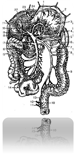
Fig. 135 Portal vein system 478 647 319 277:
1 – superior mesenteric vein 319 841 219 221
2 – stomach 898 898 478 213
3 – left gastroepiploic vein 428 816 968 989
4 – left gastric vein 319 817 298 061
5 – spleen 548 711 918 321
6 – tail of pancreas 898 429 719 482
7 – splenic vein 598 715 328 515
8 – inferior mesenteric vein 894 312 594 712
9 – descending colon 248 389 428 999
10 – rectum 598 714 898 314
11 – inferior rectal vein 594 316 814 216
12 – middle rectal vein 499 371 819 281
13 – superior rectal vein 519 371 918 991
14 – ileum 394 897 594 377
15 – ascending colon 599 213 988 713
16 – head of pancreas 319 487 914 917
17, 23 – right gastroepiploic vein 519 371 914 881
18 – portal vein 319 817 919 417
19 – gallbladder vein 419 387 918 297
20 – gall bladder 918 712 418 912
21 – duodenum 589 608 488 914
22 – liver 219 712 919 222
24 – pyloric vein 429 716 219 316
Lymphatic system 548 716 228 916
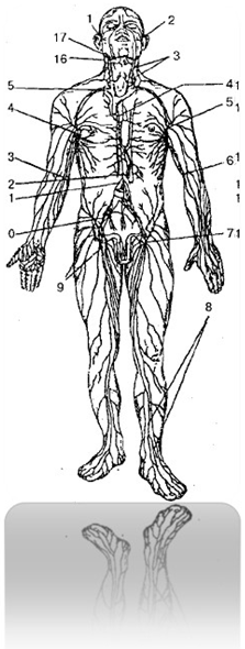
Fig. 136 Lymphatic system 548 716 228 916:
1, 2 – parotid lymph nodes 519 614 889 714
3 – cervical nodes 319 481 519 329
4 – thoracic duct 514 715 914 815
5, 14 – axillary lymph nodes 518 712 818 912
6, 13 – elbow lymph nodes 548 379 918 679
7, 9 – inguinal lymph nodes 214 387 914 297
8 – superficial lymphatic vessels of the leg
548 961 558 711
10 – iliac nodes 590 124 397 488
11 – mesenteric nodes 248 718 518 329
12 – thoracic duct cistern 519 067 819 297
15 – subclavian nodes 598 714 998 294
16 – occipital nodes 319 261 819 811
17 – submandibular nodes 548 312 819 212
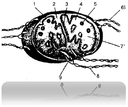
Fig. 137 Structure of the lymph node
591 148 319 888:
1 – capsule 519 848 718 949
2 – trabecula 518 716 918 317
3 – crossbar 898 749 219 317
4 – cortex 519 421 319 281
5 – follicles 898 715 984 355
6 – afferent lymphatic vessels 598 741 288 511
7 – brain substance 498 641 319 817
8 – efferent lymphatic vessels 512 789 319 489
9 – gate of the lymph node 598 681 724 918
Hematopoietic organs 498 712 818 292
ORGANS 814 317 914 817
ORGAN SYSTEMS 314 815 514 312
ORGANISM AS A SINGLE WHOLE 419 312 819 212
Content
CELLS AND TISSUES 829 379 429 841
Cells 319 078 121 942
Fabrics 898 314 988 889
Epithelial tissue 891 389 426 319
Connective tissue 719 317 918 517
Muscle tissue 514 312 814 312
Nervous tissue 718 412 518 914
NERVOUS SYSTEM 219 317 819 298
CENTRAL NERVOUS SYSTEM 291 384 074 217
Spinal cord 314 218 814 719
Brain 814 729 318 818
Medulla oblongata 514 417 814 217
Hindbrain 219 317 219 817
Pons and cerebellum 310 564 219 142
Midbrain 519 417 819 210
Diencephalon 919 213 819 223
Ventricles of the brain 989 061 714 318
Meninges of the brain 519 317 819 217
PERIPHERAL NERVOUS SYSTEM 519 555 819 915
Cranial nerves 814 212 314 812
Spinal nerves 489 218 918 217
VEGETATIVE (AUTONOMIC) NERVOUS SYSTEM
514 312 819 981
Sympathetic part of the autonomic nervous system 891 418 318 888
Parasympathetic part of the autonomic nervous system 418 217 318 918
Cytoarchitectonic fields of the cerebral cortex 219 047 819 215
Signaling systems 891 312 918 412
SENSE ORGANS 214 712 514 312
Organ of vision 219 317 989 312
Accessory organs of the eye 219 899 319 214
Organ of hearing and balance 248 712 318 222
Taste organ 419 317 819 227
Olfactory organ 914 782 214 389
Leather 519 606 901 319
BONES 214 318 214 818
BONE CONNECTIONS 814 312 214 712
Skeleton of the trunk 219 314 819 217
Skeleton of the head 231 138 918 212
Bones of the facial part of the skull 219 715 819 815
Skeleton of limbs 548 212 788 272
Bones of the upper limb 971 981 319 212
Skeleton of the free upper limb 219 317 918 817
Joints of the bones of the upper limb 248 916 319 281
Bones of the lower limb 529 531 919 811
Lower limb belt 429 712 918 222
Skeleton of the free part of the lower limb 298 321 918 557
Joints of the bones of the lower limb 918 999 000 818
MUSCULAR SYSTEM 214 712 314 222
Muscles and fascia of the trunk 519 614 819 317
Back fascia 214 718 314 888
Muscles and fascia of the head and neck 219 214 419 314
Masticatory muscles 519 314 819 214
Fascia of the head 519 718 218 314
Neck muscles 548 007 998 227
Deep Neck Muscles 819 341 919 841
Muscles and fascia of the upper limb 219 314 819 914
Muscles of the lower limb 514 311 914 527
Pelvic muscles 298 317 919 817
Muscles of the free part of the lower limb 319 715 819 555
Calf muscles 329 481 918 511
Muscles of the foot 519 371 819 511
Fascia of the lower limb 529 377 429 879
Fascia of the leg 539 427 819 677
INTERNAL ORGANS 523,000,898,111
Digestive system 541 928 741 588
Oral cavity 891 000 499 887
Oral glands 319 841 519 811
Throat 519 987 319 427
Esophagus 598 381 698 711
Stomach 898 898 478 213
Small intestine 528 317 428 717
Liver, duodenum, pancreas
219 214 319 714
Large intestine 591 488 898 217
Abdominal cavity and peritoneum 598 123 098 719
RESPIRATORY SYSTEM 598 788 428 317
Nasal cavity 428 317 818 227
Larynx 291 891 419 391
Laryngeal cartilages 529 319 489 518
Muscles of the larynx 594 318 719 214
Laryngeal cavity 581 398 421 898
Trachea and bronchi 428 714 008 914
Lungs 519 418 319 818
Pleura and mediastinum 898 315 428 188
UROGENITAL APPARATUS 898 398 412 842
Kidney 289 391 814 216
Ureters 214 312 810 008
Bladder 219 389 998 419
Urethra 329 487 948 216
Male genitalia 519 007 898 367
Female genitals 519 814 089 319
Internal female genital organs 419 219 808 319
ENDOCRINE GLANDS 889 314 219 798
Pituitary and pineal gland 214 318 908 210
Pituitary gland 317 218 219 819
Pineal body (Epiphysis) 489 641 399 042
Thyroid and parathyroid glands 219 318 219 471
Thymus gland 481 914 319 814
Thyroid gland 829 319 409 819
Parathyroid glands 219 319 895 219
Adrenal gland 891 418 712 319
Endocrine pancreas 918 712 818 229
Endocrine part of the sex glands 519 318 914 019
Regulation of endocrine glands 519 317 219 416
CARDIOVASCULAR SYSTEM 214 700 819 891
Arteries, veins and capillaries 219 387 919 887
Heart 918 749 328 081
Conduction system of the heart 989 808 884 318
Blood supply and innervation of the heart 891 318 910 488
Vessels of the pulmonary circulation 898 714 988 569
Pulmonary veins 309 428 519 421
Vessels of the systemic circulation 598 716 588 317
Branches of the aortic arch 298 714 319 814
Branches of the thoracic aorta 548 617 908 614
Branches of the abdominal aorta 548 711 488 211
Veins of the systemic circulation 419 641 819 317
Veins of the heart 891 428 319 298
Superior vena cava system 898 317 419 217
Veins of the head and neck 598 716 319 816
Inferior vena cava system 394 617 014 809
Portal vein system 478 647 319 277
Lymphatic system 548 716 228 916
Hematopoietic organs 498 712 818 292
ORGANS 814 317 914 817
ORGAN SYSTEMS 314 815 514 312
ORGANISM AS A SINGLE WHOLE 419 312 819 212